
Medical image denoising and enhancing processing method

A technology of medical images and processing methods, applied in image enhancement, image data processing, instruments, etc., can solve problems such as hindering applications

Active Publication Date: 2011-07-27
深圳蓝影医学科技股份有限公司
View PDF1 Cites 21 Cited by
  • Summary
  • Abstract
  • Description
  • Claims
  • Application Information

AI Technical Summary

Problems solved by technology

[0017] In the practical application of anisotropic filtering, because multiple diffusions are required to achieve the target effect, speed becomes a major constraint hindering its application.

Method used

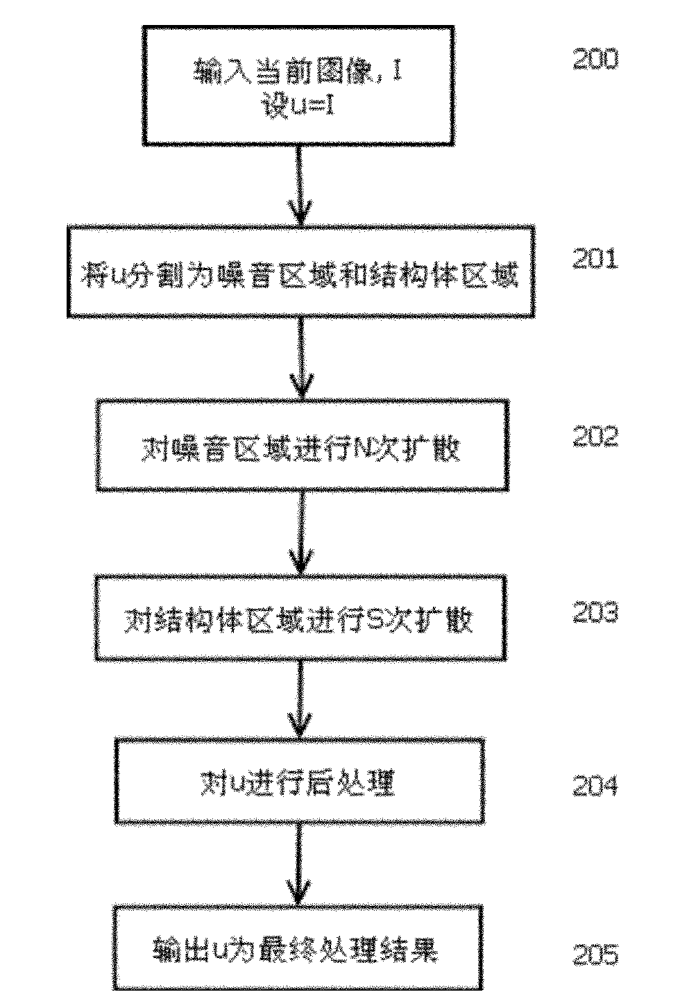

Image

  • Medical image denoising and enhancing processing method
  • Medical image denoising and enhancing processing method
  • Medical image denoising and enhancing processing method

Examples


Embodiment Construction

[0063] Below according to accompanying drawing and embodiment the present invention will be described in further detail:

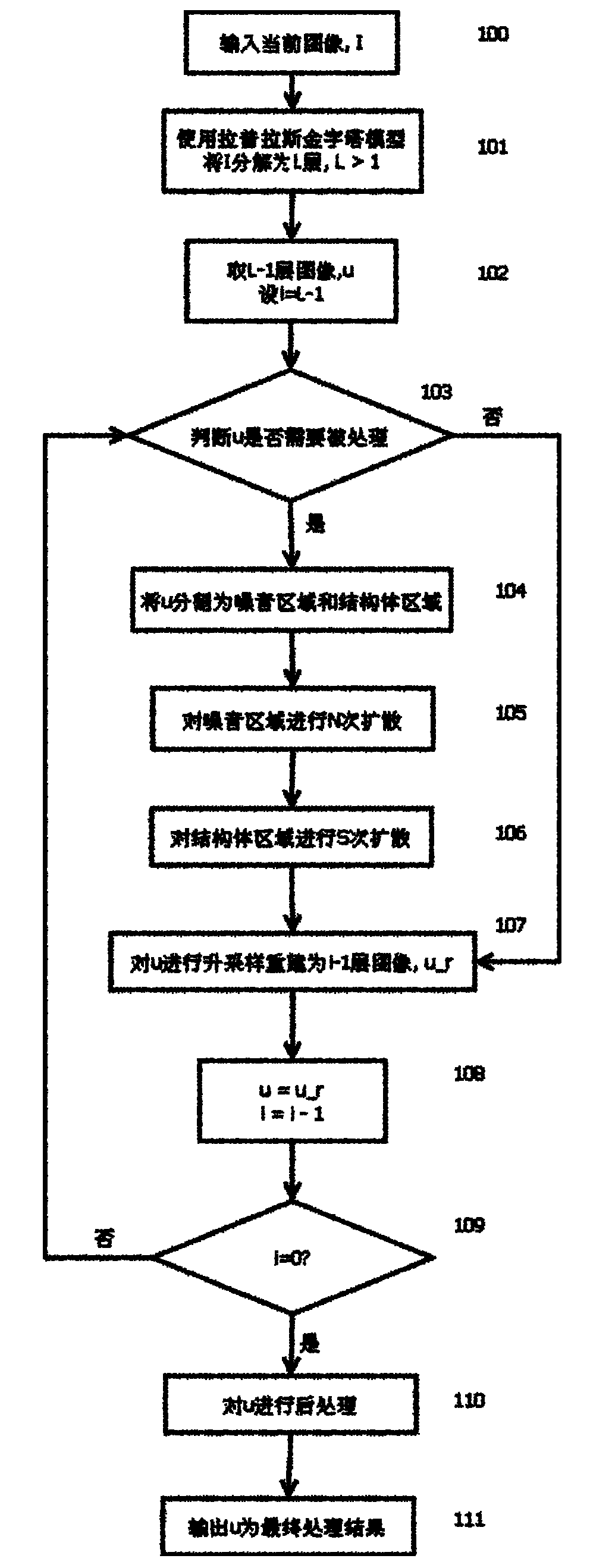
[0064] attached figure 1 It is a flow chart of the image noise reduction and enhancement method in a preferred embodiment of the invention. This processing method is suitable for noise reduction and enhancement of most medical images, such as ultrasound B-mode images, X-ray fluoroscopy images, magnetic resonance images, and CT images etc. The present invention mainly uses the diffusion tensor to implement different intensities of diffusion filtering on the noise area and the structure area, and uses multiple scales to increase the running speed and reduce the artifacts caused by the loss of low-frequency signals to improve the image granularity. The specific process of the program is as follows.

[0065] Starting from step 100, a medical image, I, is first acquired from an image acquisition device or an image storage.

[0066] In step 101, use the Lapla...


PUM


Abstract

The invention provides a real-time medical video image denoising method, which is applied to a real-time acquired or stored medical video image sequence. Time-space domain recursive filtering processing is performed on each frame of video image by utilizing high-degree correlation of two adjacent images, and the size of a filter window can be adaptively adjusted according to the noise level. In the method, a time-domain motion compensation method - and a space-domain denoising enhancement algorithm are combined; aimed at the current pixel of the image, the motion compensation method and a morphological operator are applied to detect a motion area, a noise area and a background area, and the overall noise level of the image is estimated in the noise area.

Description

technical field [0001] The invention relates to a method for noise reduction and enhancement of acquired medical images. Background technique [0002] There is usually a lot of speckle noise in medical ultrasound B-mode images, which will bring about a significant decline in the quality of ultrasound images and cover up some important tissue lesions, which brings difficulties to doctors' diagnosis and identification of certain specific diseases. There are great difficulties, and there is a risk of missed diagnosis and misdiagnosis. [0003] The filtering method is usually used to suppress the high-frequency part of the signal, and the useful edge information will be removed while filtering out the noise. Over-smoothing; As a result, some filtering algorithms based on edge preservation have been developed rapidly, such as anisotropic diffusion, BM3D, and nonlocal means and other medical image denoising techniques. This provides a huge space for improving the quality of medi...

Claims


Application Information

Patent Timeline
Patent Type & Authority Applications(China)
IPC IPC(8): G06T5/00
Inventor 陆汇海张羽田镇魁潘梁亮
Owner 深圳蓝影医学科技股份有限公司
Learn More