Method for effectively culturing tumor infiltrating lymphocytes (TILs)
A lymphocyte and tumor infiltration technology, applied in anti-tumor drugs, animal cells, vertebrate cells, etc., can solve the problems of lack of specificity of TIL cells, slow cell proliferation, and limited clinical efficacy, so as to reduce the burden of hospitalization and reduce hospitalization. The effect of shortening time and enhancing clinical efficacy
- Summary
- Abstract
- Description
- Claims
- Application Information
AI Technical Summary
Problems solved by technology
Method used
Image
Examples
Embodiment 1
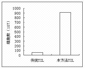
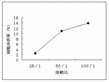
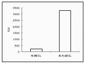
[0027] The patient, male, 70 years old, had right lung cancer and a large amount of pleural effusion. Transfer 800ml of pleural effusion to a centrifuge tube, centrifuge at 1500rpm × 8min, take the supernatant of pleural effusion, mix the sediment with serum-free medium, separate the cells with lymphocyte separation medium and centrifuge 800g×20min. Aspirate the buffy coat, centrifuge at 2000rpm×10min, add serum-free medium, centrifuge at 1600rpm×8min, discard the supernatant to obtain mononuclear cells. Adjust the number of cells to 1.5 x 10 6 / ml, inoculate with RetroNectin250 μl, CD 3 Antibody 100μl Coated 175cm 2 In a culture bottle, placed in a saturated humidity of 90%, a temperature of 37°C, and CO 2 5.0% cultured in the incubator. After culturing for 24 hours, add IL-2 1000U / ml, and continue to store at 90% saturated humidity, temperature 37°C, CO 2 5.0% incubator culture. On the third day, the TIL cells were subcultured with serum-free medium containing 1000 U...
Embodiment 2
[0032] The patient, male, 65 years old, had left lung cancer. The surgical tissue of the lung was taken and placed in a sterile tank, and the tumor specimen was washed 2 or 3 times with sterile saline containing gentamicin 100-160 IU / ml. Add 10-20ml of culture medium to a 10cm diameter petri dish, put a piece of 200 mesh metal mesh, cut the tumor tissue into small pieces and place on the metal mesh. Crush the tumor tissue block with a 20ML syringe core, discard the metal mesh, collect the serum-free medium in the culture dish (cells flow into the medium to form a single-cell suspension), add the cell suspension to 20ml serum-free medium, 300RPM×3min. Take the supernatant, 1500 RPM × 6min, the sedimentation is the single cell layer. Add serum-free medium and mix well, separate the cells with lymphocyte separation medium and centrifuge at 800g×20min. Aspirate the buffy coat, centrifuge at 2000rpm×10min, add serum-free medium, centrifuge at 1600rpm×8min, discard the supernatant...
Embodiment 3
[0034] The patient, female, 68 years old, had multiple bone metastases from gastric adenocarcinoma and a large amount of ascites. Aseptically draw 650ml of pleural fluid. Transfer 650ml of pleural effusion to a 50Ml centrifuge tube, centrifuge at 1800rpm × 6min, take the supernatant of pleural effusion, mix the sediment with serum-free medium, separate cells with lymphocyte separation medium, and centrifuge at 800g × 22min. Aspirate the buffy coat, centrifuge at 2000rpm×10min, add serum-free medium, centrifuge at 1500rpm×8min, discard the supernatant to obtain mononuclear cells. Adjust the number of cells to 2.0×10 6 / ml, inoculate with RetroNectin250μl, CD with serum-free medium containing 0.01ml / ml of pleural fluid supernatant 3 Antibody 100μl Coated 175cm 2 In a culture bottle, placed in a saturated humidity of 90%, a temperature of 37°C, and CO 2 5.0% cultured in the incubator. After culturing for 24 hours, add IL-2 1500U / ml to the cells, and continue to store at 90%...
PUM
 Abstract
Description
Claims
Application Information
 - R&D
- Intellectual Property
- Life Sciences
- Materials
- Tech Scout
- Unparalleled Data Quality
- Higher Quality Content
- 60% Fewer Hallucinations
Browse by: Latest US Patents, China's latest patents, Technical Efficacy Thesaurus, Application Domain, Technology Topic, Popular Technical Reports.
© 2025 PatSnap. All rights reserved.Legal|Privacy policy|Modern Slavery Act Transparency Statement|Sitemap|About US| Contact US: help@patsnap.com


