
Method for culturing human embryo stem cell by using umbilical cord source mesenchymal stem cell

A technology of human embryonic stem cells and mesenchymal stem cells, applied in the field of culturing human embryonic stem cells using umbilical cord-derived mesenchymal stem cells, can solve problems such as difficulties in clinical research and application, achieve small complications and risks, maintain stability, and maintain undifferentiated growth Effect

Inactive Publication Date: 2011-11-23
PEKING UNIV THIRD HOSPITAL
View PDF4 Cites 5 Cited by
  • Summary
  • Abstract
  • Description
  • Claims
  • Application Information

AI Technical Summary

Problems solved by technology

[0004] But for hESCs, the initial hESCs line was established with mouse embryonic fibroblast cells (MEF) as the feeder layer. Due to the necessary feeder layer for the culture system and the animal-derived components of the culture system These problems make the clinical research and future application of cells face many difficulties

Method used


Image

  • Method for culturing human embryo stem cell by using umbilical cord source mesenchymal stem cell
  • Method for culturing human embryo stem cell by using umbilical cord source mesenchymal stem cell
  • Method for culturing human embryo stem cell by using umbilical cord source mesenchymal stem cell

Examples


Embodiment 1

[0079] Embodiment 1, the preparation of experimental cell

[0080] 1. Primary culture, passage, cryopreservation and recovery of human umbilical cord mesenchymal stem cells (HUCMSCs)

[0081] 1. Primary culture of HUCMSCs

[0082] (1) Aseptically take the isolated umbilical cord 10cm of healthy (no infectious disease, serological examination is negative for HBV, HCV, HIV and syphilis) caesarean section fetus, and place it in a 50ml centrifuge tube, which is put into sterile PBS in advance; the umbilical cord The acquisition was obtained with the informed consent of the pregnant woman and her family members.

[0083] (2) In the ultra-clean bench, fully rinse the outer surface of the umbilical cord with PBS containing 1× penicillin-streptomycin, fully wash off the blood, and then put the umbilical cord into a glass dish for operation;

[0084] (3) Use tissue scissors to carefully remove two umbilical veins and one umbilical artery, and cut the remaining umbilical cord intersti...

Embodiment 2

[0163] Example 2, Proliferation and Culture of Human Embryonic Stem Cells

[0164] 1. Proliferation and culture of human embryonic stem cells by the method of the present invention

[0165] (1) Preparation of HUCMSCs feeder layer

[0166] (1) Take HUCMSCs from P3 to P13, wait until they reach the logarithmic growth phase, discard the medium, and add a medium containing 10 μg / ml mitomycin C (that is, the concentration of mitomycin C in the medium is 10 μg / ml), placed in the incubator for 3 hours;

[0167] (2) Treat the petri dish with 0.1% gelatin (add gelatin, shake to cover the bottom of the dish with gelatin, then place at 37°C for half an hour), discard the gelatin, and place at room temperature until the gelatin at the bottom of the dish is dry.

[0168] (3) Discard the medium containing mitomycin C, add 2-3mL PBS, shake gently, discard the PBS, and wash 3-5 times in this way (must be washed more than 3 times to completely wash away the remaining mitomycin Mitomycin C, ...

Embodiment 3

[0186] Example 3. Identification of hESCs using HUCMSCs and MEFs as feeder cells

[0187] 1. Alkaline phosphatase (AKP) staining

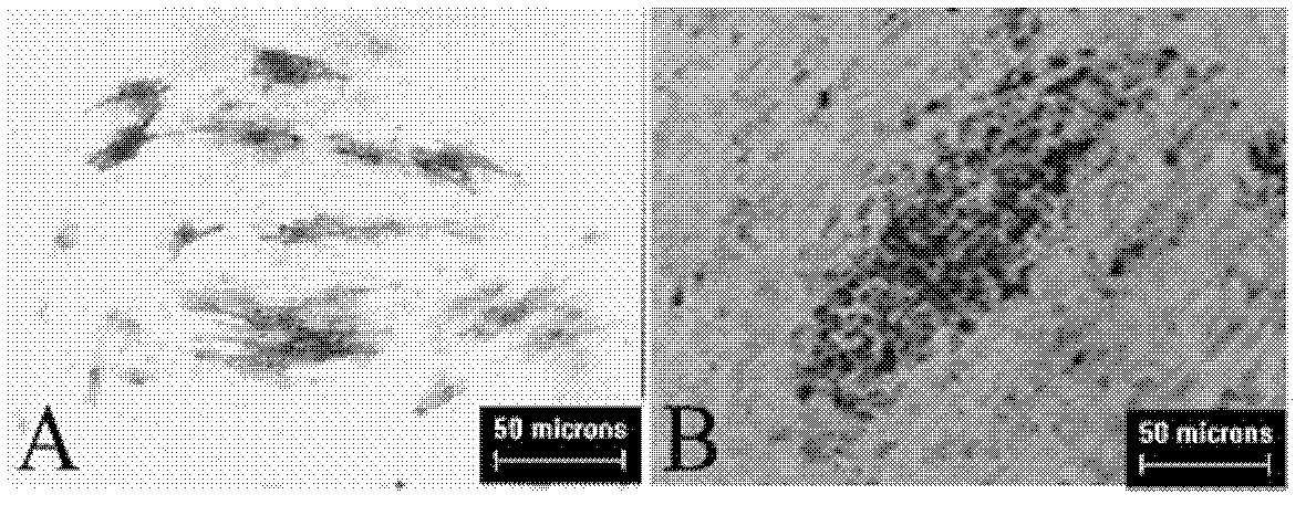
[0188] Positive alkaline phosphatase staining is one of the indicators of hESCs pluripotency. hESCs at passage 30 were stained and observed using the Millipore Alkaline Phosphatase Detection Kit. Refer to the manual for specific steps. Observation and photography under a microscope.

[0189] Identified by alkaline phosphatase staining, hESCs alkaline phosphatase staining on the HUCMSCs feeder layer ( Figure 7 A) The alkaline phosphatase staining of hESCs with MEF as the feeder layer was positive ( Figure 7 B).

[0190] 2. Immunofluorescence detection of specific antigen expression

[0191] (1) After hESCs were cultured on HUCMSCs feeder layer and MEF feeder layer for 30 passages, appropriate amount of cells were digested and inoculated in a 24-well plate with feeder layer cells.

[0192] (2) After 3 days, fix with 4% paraformaldehyde at ro...


PUM


Abstract

The invention discloses application of an umbilical cord source mesenchymal stem cell and a method for culturing a human embryo stem cell. An enrichment culturing method for a human embryo stem cell, provided by the invention, comprises the following step of: performing enrichment culturing on an isolated human embryo stem cell by taking an isolated umbilical cord mesenchymal stem cell as a feeder layer to obtain proliferative human embryo stem cells. As proved by the invention, the umbilical cord source mesenchymal stem cell is an ideal anthropogenic feeder layer source cell for maintaining the human embryo stem cell in an undifferentiated growing status, and a novel thought train is provided for the problems caused in the clinical application of the human embryo stem cell.

Description

technical field [0001] The invention relates to the application of umbilical cord-derived mesenchymal stem cells and the culture method of human embryonic stem cells. Background technique [0002] Human-derived stem cells refer to cell populations with self-renewal, proliferation and multi-directional differentiation potentials in the growth and development of human individuals. They are divided into embryonic stem cells (human embryonic stem cells, hESCs), adult stem cells, and transgenic The technology converts adult skin fibroblasts into induced pluripotent stem cells (iPS) with multiple differentiation potential. [0003] hESCs were first isolated from the inner cell mass (ICM) of human blastocysts, and their most important characteristics are: (1) high self-replication ability and near unlimited proliferation ability in vitro; (2) developmental Totipotency, ES cells can differentiate into all tissue cells of a complete individual, providing sufficient cell sources for ...

Claims


Application Information

Patent Timeline
Patent Type & Authority Applications(China)
IPC IPC(8): C12N5/0735
Inventor 张纯徐永胜王薇张绍敏俞海燕刘峰
Owner PEKING UNIV THIRD HOSPITAL
Learn More