Three-dimensional imaging device for retina

A three-dimensional imaging and retinal technology, which is used in medical science, ophthalmoscope, and eye testing equipment. The effect of eliminating non-uniformity, eliminating stray light

A three-dimensional imaging and retinal technology, which is used in medical science, ophthalmoscope, and eye testing equipment. The effect of eliminating non-uniformity, eliminating stray light

CN102525406AInactive Publication Date: 2012-07-04SUZHOU MICROCLEAR MEDICAL INSTR

Image

  • Three-dimensional imaging device for retina
  • Three-dimensional imaging device for retina
  • Three-dimensional imaging device for retina

Examples


Embodiment Construction

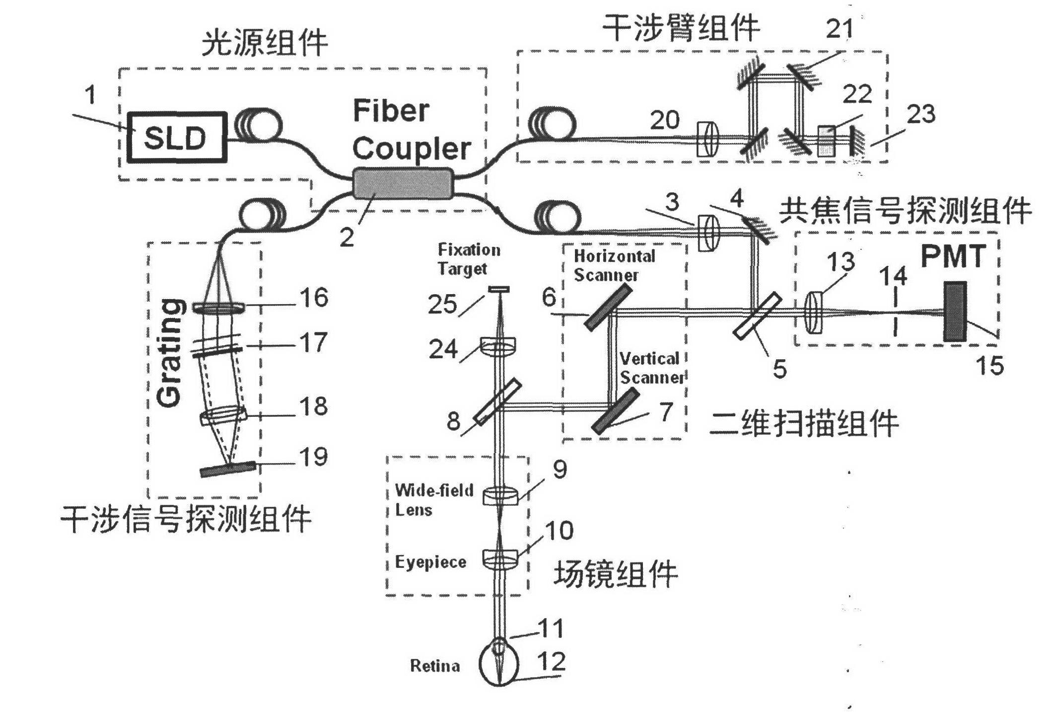
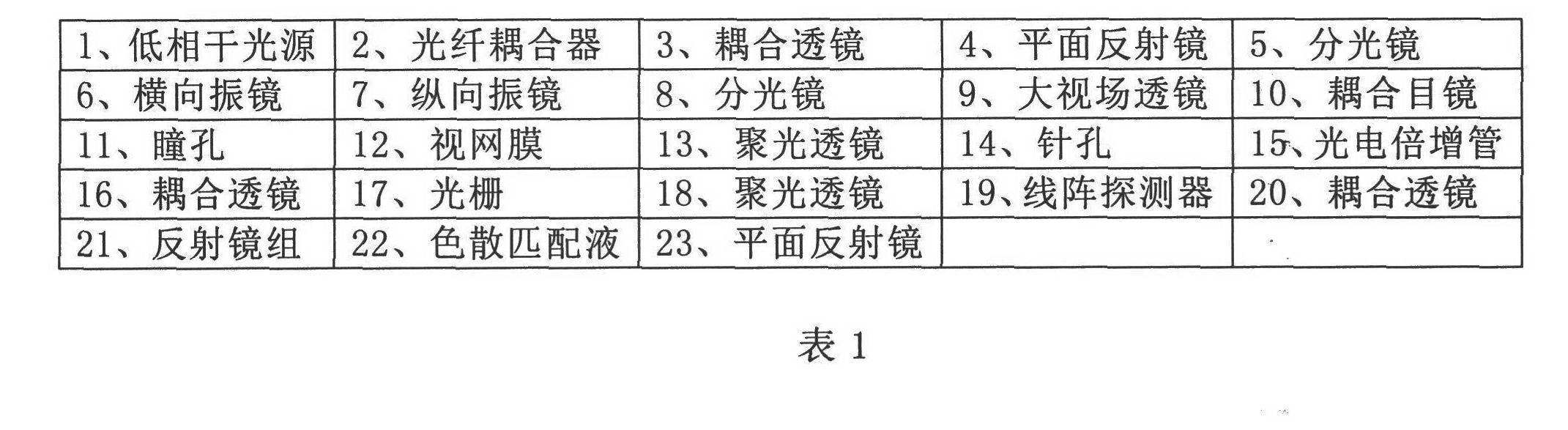
[0022] According to the manual attached figure 1 , to how to implement the function of a kind of retina three-dimensional imaging device that the present invention proposes, detailed introduction is as follows:

[0023] 1. The low-coherence light source SLD (1) of the light source component is connected to the multi-channel fiber coupler (2) through the fiber port. The fiber coupler (2) has two output ports, one fiber output to the interference arm component, and the coupling lens (20) Collimate, after collimation, it becomes parallel light and enters the reflector assembly (21) and dispersion matching liquid (22), and is reflected by the reflector (23), and the original optical path of the optical signal returns to the fiber coupler (2) , and then enter the interference signal detection component.

[0024] 2. The other beam after passing through the fiber coupler (2) is collimated into parallel light by the coupling lens (3), and then reflected into the two-dimensional scann...


PUM


Abstract

The invention discloses a three-dimensional imaging device for a retina. The device consists of a light source component, an interference arm component, a two-dimensional scanning component, a field lens component, a confocal signal detection component and an interference signal detection component. The three-dimensional imaging device for the retina finishes scanning on the retina of a human eye by using the two-dimensional scanning component, extracts a surface image of the retina by using the confocal signal detection component and depth information of the retina by using the interference signal detection component, and jointly finishes the reconstruction of a three-dimension image of the retina through a confocal signal and an interference signal; the system is compact by adopting the design of optical fiber access and an optical fiber coupler; the influence of curvature of field and aberration in the imaging process is reduced through the design of a field lens so as to acquire a large-field-of-view three-dimensional image of the retina of the human eye; and the three-dimensional imaging device for the retina which is compactly designed and is high in imaging resolution and large in imaging field of view is realized, so that the imaging effect of the traditional fundus imaging instrument is greatly improved.

Description

technical field [0001] The invention relates to an ophthalmic imaging device, in particular to a three-dimensional retinal imaging device, which can scan and image different areas and thicknesses of the retina within a large field of view, reconstruct the three-dimensional retinal image and output the observation results via video, Provide scientific reference for the diagnosis of retinal diseases. Background technique [0002] Confocal scanning imaging and optical coherence tomography have been widely and maturely applied in the field of biological histology imaging (Webb RH, Hughes GW. Scanning Laser Ophthalmoscope. Biomedical Engineering, IEEE Transactions on.1981, BME-28 (7 ): 488-92.); A.F.Fercher et al earlier proposed to measure the length of the eye axis by interferometric imaging (A.F.Fercher et.Al "Eye-length measurement by interferometry with partially coherent light", Optics Letters.13, 186 -188 (1998)), and then David Huang et al. did more research on the devel...

Claims


Application Information

Patent Timeline
04 Jul 2012
Publication
CN102525406A
IPC
A61B3/12; A61B3/15
Inventors
李超宏