Living cell laser scanning co-focusing microscope imaging system

A technology of confocal microscopy and confocal microscopy, applied in microscopes, optics, optical components, etc., can solve the problems of reducing the signal-to-noise ratio, etc., and achieve the effect of high precision, reducing the degree of phototoxicity and photobleaching, and compact structure

Inactive Publication Date: 2012-08-01
FUJIAN NORMAL UNIV
View PDF12 Cites 24 Cited by
  • Summary
  • Abstract
  • Description
  • Claims
  • Application Information

AI Technical Summary

Problems solved by technology

Although the degree of phototoxicity and photobleaching can be reduced by reducing the li

Method used


Image

  • Living cell laser scanning co-focusing microscope imaging system

Examples


Embodiment Construction

[0012] The present invention will be further elaborated below in conjunction with the accompanying drawings and embodiments.

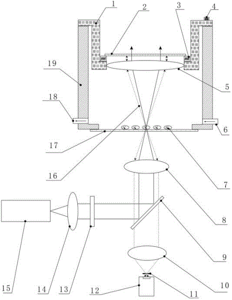
[0013] refer to figure 1 , a live cell laser scanning confocal microscope imaging system, including a laser scanning confocal microscope and a fluorescence signal acquisition device, the fluorescence signal acquisition device includes a culture dish 19 and a converging lens 5 and a reflective type arranged in the culture dish 19. Narrow-band filter 2; the bottom of the culture dish 19 is provided with a culture dish bottom plate 17 for placing the cells to be tested 7; The cell to be measured 7 on the 17 is located on the focal plane of the condensing lens 5; the reflective narrow-band filter 2 is located directly above the condensing lens 5; the objective lens 8 of the laser scanning confocal microscope is located directly on the bottom plate of the culture dish 17 below.

[0014] In this embodiment, the reflective narrow-band filter 2 is disposed o...


PUM


Abstract

The invention relates to a living cell laser scanning co-focusing microscope imaging system, which is characterized by comprising a laser scanning co-focusing microscope and a fluorescent signal collecting device, wherein the fluorescent signal collecting device comprises a culture dish, a converging lens and a reflecting type narrow-band light filter, the converging lens and the reflecting type narrow-band light filter are arranged in the culture dish, the bottom of the culture dish is provided with a culture dish bottom plate used for placing cells to be tested, the converging lens is fixedly arranged on the culture dish through a lens clamping frame, the cells to be tested on the culture dish bottom plate are positioned on the focal plane of the converging lens, the reflecting type narrow-band light filter is positioned right above the converging lens, and an objective lens of the laser scanning co-focusing microscope is positioned right under the culture dish bottom plate. The living cell laser scanning co-focusing microscope imaging system has the advantages that the structure is compact, the precision is high, the fluorescent signal collection efficiency of the laser scanning co-focusing microscope can be effectively improved, the phototoxicity and the light bleaching degree are reduced, and a technical means is provided for obtaining reliable experiment results.

Description

technical field [0001] The invention relates to a live cell laser scanning confocal microscope imaging system. Background technique [0002] Live cell optical imaging technology is an important means to study the physiological and pathological mechanisms at the cellular level. In live cell optical imaging, phototoxicity and photobleaching are two important factors, which play a crucial role in the accuracy of experimental results, especially for long-term observation of cell physiology and pathology. Phototoxicity affects the viability of cells and indirectly affects the experimental results, while photobleaching can affect the obtained fluorescence signal and has a direct impact on the experimental results. Therefore, minimizing the degree of phototoxicity and photobleaching has become a common concern in live-cell optical imaging techniques. Although phototoxicity and photobleaching can be mitigated by reducing the laser irradiation dose, the signal-to-noise ratio will a...

Claims


Application Information

Patent Timeline
IPC IPC(8): G01N21/64G02B21/36
Inventor 黄义梅谢树森杨洪钦李晖王瑜华
Owner FUJIAN NORMAL UNIV
Why Eureka
Try Eureka