Diagnosis and treat integral confocal cystoscope system

A cystoscopy and confocal technology, applied in cystoscopy, diagnosis, endoscopy, etc., can solve the problems of increasing patient pain, wasting operation time, and unfavorable operation, so as to save operation time, improve accuracy and safety sexual effect

Inactive Publication Date: 2014-11-05
GUANGZHOU BAODAN MEDICAL INSTR TECH
View PDF5 Cites 0 Cited by
  • Summary
  • Abstract
  • Description
  • Claims
  • Application Information

AI Technical Summary

Problems solved by technology

The doctor passes the cystoscope through the patient's urethra, and can observe the real situation in the bladder cavity through the optical system of the endoscope, and pass in surgical instruments to treat bladder lesions. For suspicious lesions such as tumors and inflammations in the bladder cavity or mucous membrane , Doctors often need to access surgical instruments such as biopsy forceps to obtain tissues and send them for inspection. The process of sending for inspection takes a long time and often delays the precious treatment time of patients
After the lesion is diagnosed, it is necessary to frequently access different types of instruments, such as laser knife probe for resection of the lesion, microwave knife probe for microwave treatment of the lesion, etc., which increases the pain of the patient and wastes a lot of operation time, and is not conducive to the smooth operation conduct

Method used


Image

  • Diagnosis and treat integral confocal cystoscope system
  • Diagnosis and treat integral confocal cystoscope system
  • Diagnosis and treat integral confocal cystoscope system

Examples


Embodiment 1

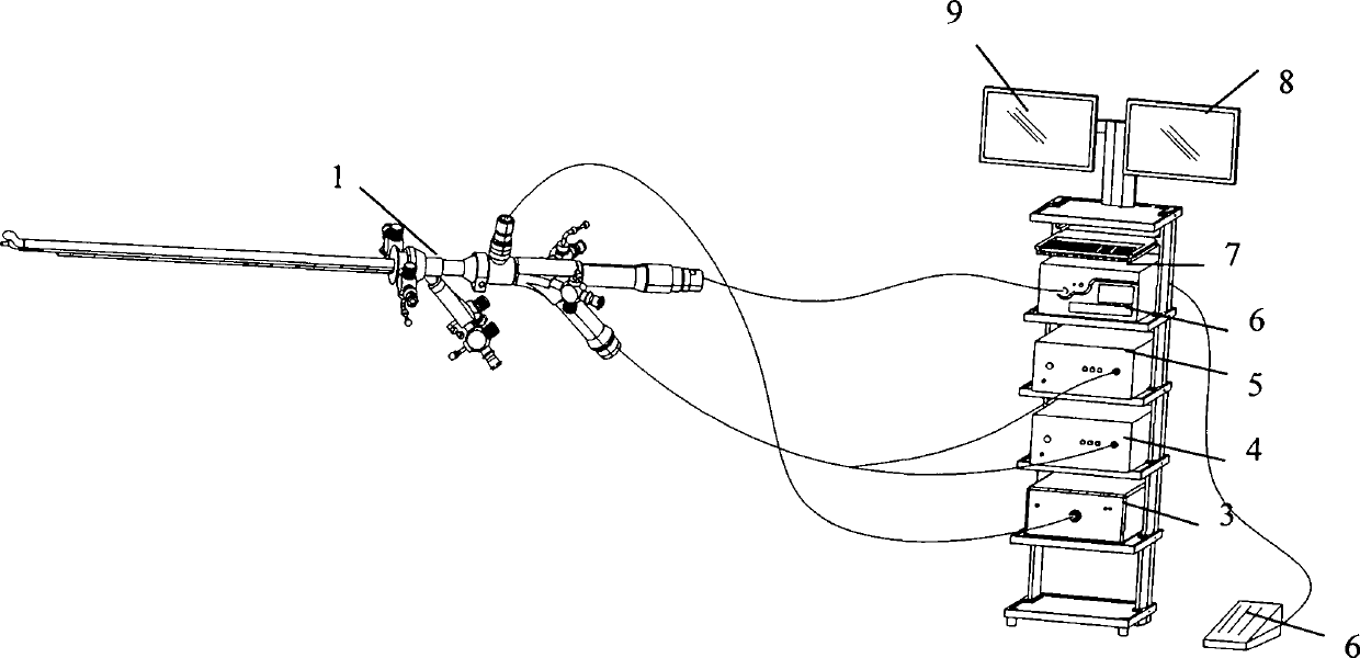
[0028] Such as figure 1 As shown, the diagnosis and treatment integrated confocal cystoscope system of the present invention includes a rigid cystoscope 1, a cold light source host 3 connected to the rigid cystoscope, a camera host 4, a confocal laser scanning microscope system host 5, Laser knife system host 6, keyboard 7 and monitors 8,9.

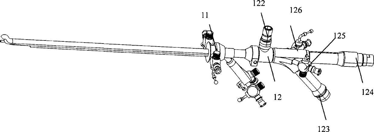
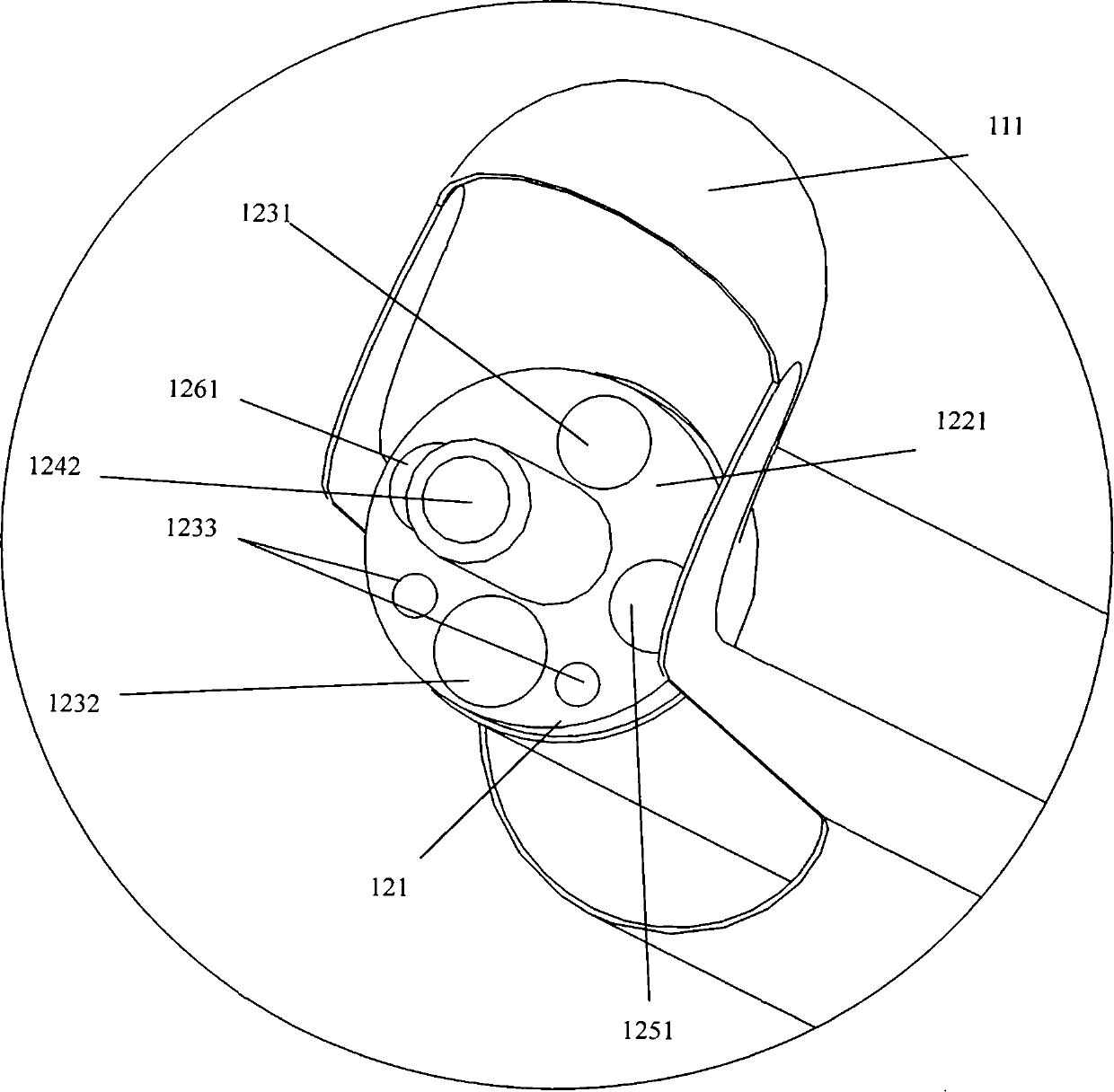
[0029] Such as figure 2 As shown, the rigid cystoscope 1 of the present invention includes two parts: a cystoscope sheath 11 and a cystoscope main body 12 . The maximum outer diameter of the cystoscope sheath 11 is less than or equal to 12mm, and the tip 111 of the cystoscope sheath 11 is designed to be blunt to avoid damage to urethral tissue. The structure of the cystoscope main body 12 includes a rigid endoscope end 121 , a cold light source connector 122 , an image data output 123 , a laser knife control interface 124 , a water inlet channel 125 and a water outlet channel 126 and the like. The image data output terminal 123 is res...

Embodiment 2

[0033] This embodiment is basically the same as the first embodiment above, the difference is:

[0034] Such as Figure 5 As shown, the diagnosis and treatment integrated confocal cystoscope system of the present invention includes a rigid cystoscope 2, a cold light source host 3 connected to the rigid cystoscope, a camera host 4, a confocal laser scanning microscope system host 5, Microwave knife system host 10, keyboard 7 and monitors 8,9.

[0035] Figure 6 As shown, the rigid cystoscope 2 includes two parts: a cystoscope sheath 21 and a cystoscope main body 22 . The maximum outer diameter of the cystoscope sheath 21 is less than or equal to 12 mm, and the tip 211 of the cystoscope sheath 21 is designed to be blunt to avoid damage to urethral tissue. The structure of the cystoscope main body 22 includes a rigid endoscope end 221 , a cold light source connector 222 , an image data output 223 , a microwave knife control interface 224 , a water inlet channel 225 and a water...


PUM


Abstract

The invention belongs to the field of medical machinery, and particularly relates to a diagnosis and treat integral confocal cystoscope system which comprises a hard cystoscope, wherein a treatment unit is arranged at the end part of a hard endoscope of the hard cystoscope, and a host machine of a treatment system is connected onto the hard cystoscope and correspondingly matched with the treatment device for use. The diagnosis and treat integral confocal cystoscope system is organically combined with a laser scalpel system or a microwave scalpel system through the hard cystoscope and a confocal laser scanning microscope system. The clinical application of the diagnosis and treat integral confocal cystoscope system can realize the effect of simultaneous diagnosis and treatment, and also can solve the problems of two aspects of patient diagnosis and treatment, the endoscope is prevented from being frequently changed, a large amount of operation time is saved, the pain of a patient is relieved, furthermore, the operation accuracy and safety are improved, and an unexpected effect is obtained.

Description

technical field [0001] The invention belongs to the field of medical equipment, and in particular relates to a confocal cystoscope system integrating diagnosis and treatment for bladder disease surgery. Background technique [0002] At present, cystoscopy is widely used in the diagnosis and treatment of urological surgery, and plays an important role in the treatment of bladder stones and bladder-related lesions. The doctor passes the cystoscope through the patient's urethra, and can observe the real situation in the bladder cavity through the optical system of the endoscope, and pass in surgical instruments to treat bladder lesions. For suspicious lesions such as tumors and inflammations in the bladder cavity or mucous membrane , Doctors often need to access surgical instruments such as biopsy forceps to obtain tissues and send them for inspection. The process of sending them for inspection takes a long time and often delays the precious treatment time of patients. After t...

Claims


Application Information

Patent Timeline
Patent Type & Authority Patents(China)
IPC IPC(8): A61B1/307A61B1/04A61B1/07A61B18/22A61B18/18
Inventor 乔铁
Owner GUANGZHOU BAODAN MEDICAL INSTR TECH
Why Eureka
Try Eureka