Quantitative biomolecule detection method

A quantitative detection method and technology of biomolecules, applied in the field of quantitative detection of biomolecules, can solve the problems of low sensitivity, cumbersome steps, and high cost, and achieve the effects of improved detection sensitivity, mild reaction conditions, and reduced detection costs

Inactive Publication Date: 2012-09-05
HEBEI UNIVERSITY
View PDF1 Cites 14 Cited by
  • Summary
  • Abstract
  • Description
  • Claims
  • Application Information

AI Technical Summary

Problems solved by technology

However, there is still a lack of sensitive and effective means for how to analyze the HCR products and then give the quantitative information of the initial target molecules.
Currently, the reported methods for detecting HCR products mainly include gel electrophoresis and fluorescent labeling methods. The gel electrophoresis detection method has low sensitivity and cannot meet the analysis requirements of trace biomolecules; Tan et al. (Angew.Chem.Int.Ed. , 2011, 50, 401-404) were labeled with Pyrene fluorescent molecules at the two ends of the H1 and H2 probes, and Pyrene dimers would be formed after the HCR reaction, whose fluo...

Method used


Image

  • Quantitative biomolecule detection method
  • Quantitative biomolecule detection method
  • Quantitative biomolecule detection method

Examples


Embodiment 1

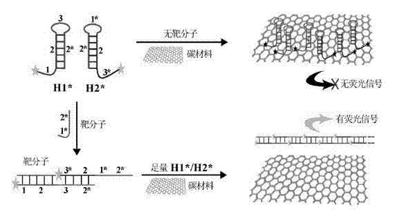
[0033] (a) Using let-7a microRNA as the target biomolecule (purchased from Dalian Bao Biological Co., Ltd.), according to its sequence (UGAGGUAGUAGGUUGUAUAGUU), design FAM fluorescently labeled H1* and H2* probes according to conventional methods, and the designed H1*, The H2* probe sequences are: H1*: AGTAGGTTGTATAGTTCAAAGTAACTATACAACCTACTACCTCA-FAM; H2*: FAM-ACTTTGAACTATACAACCTACTTGAGGTAGTAGGTTGTATAGTT.

[0034] (b) Place H1* and H2* monomer solutions with a concentration of 2 μM on a PCR instrument and heat at 95°C for 2 minutes, and then place them at room temperature in the dark for 1 hour to fully close the stem-loop structure;

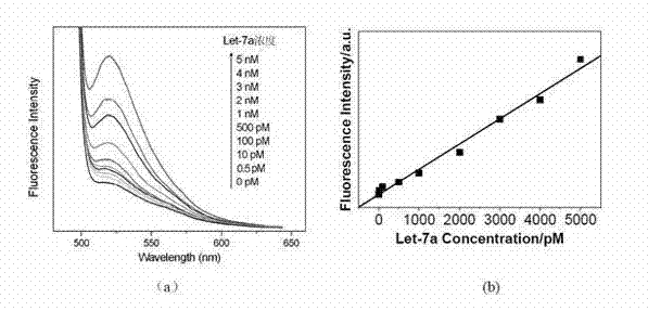
[0035](c) Take 10 RNase-free centrifuge tubes, add different concentrations of target let-7a microRNA to each tube, so that the final concentration of let-7a in each tube is 0pM, 0.5pM, 10pM, 100pM, 500pM, 1nM , 2nM, 3nM, 4nM, 5nM, and then add H1* and H2* with a final concentration of 50nM to each centrifuge tube, a certain volume of buffer sol...

Embodiment 2

[0040] (a) Let-7a mi croRNA was used as the target detection molecule, and FAM fluorescently labeled H1* and H2* probes were designed according to its sequence (UGAGGUAGUAGGUUGUAUAGUU), and the sequences were: H1*: AGTAGGTTGTATAGTTCAAAGTAACTATAC AACCTACTACCTCA-FAM; H2*: FAM -ACTTTGAACTATACAACCTACTTGAGGTAGTAGGTTGTATAGTT;

[0041] (b) Place H1* and H2* monomer solutions with a concentration of 2 μM on a PCR instrument and heat at 95°C for 2 minutes, and then place them at room temperature in the dark for 1 hour to fully close the stem-loop structure;

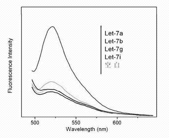
[0042] (c) Take 4 RNase-free centrifuge tubes, add let-7a, let-7b, let-7g, and let-7i at the same concentration (3nM) to each tube, and take another centrifuge tube as a blank control at the same time ; Then each centrifuge tube was added with a final concentration of 50nM H1* and H2*, a certain volume of buffer solution and water, so that the final reaction medium in each tube was 200 μL volume of SSP buffer (50mM Na 2 HPO 4 , ...

Embodiment 4

[0046] (a) Take a randomly selected sequence DNA fragment (selected from Proc.Natl.Acad.Sci.USA, 2004, 15275-15278) as the target detection molecule, and design FAM-labeled H1* and H2 according to its sequence (AGTCTAGGATTCGGCGTGGGTTAA) *Probes, the sequences are: H1*: FAM-TTAACCCACGCCGAATCCTAGACTCAAAGTAGTCTAGGATTCGGCGTG; H2*: AGTCTAGGATTCGGCGTGGGTTAACACGCCGAATCCTAGACTACTTTG-FAM;

[0047] (b) Place H1* and H2* monomer solutions with a concentration of 2 μM on a PCR instrument and heat at 95°C for 2 minutes, and then place them at room temperature in the dark for 1 hour to fully close the stem-loop structure;

[0048] (c) Take 10 centrifuge tubes, add different concentrations of target DNA molecules into each tube, so that the final concentrations are 0, 0.5pM, 10pM, 100pM, 500pM, 1nM, 2nM, 3nM, 4nM, 5nM, and then Add H1* and H2* with a final concentration of 50nM, a certain volume of buffer solution and water to each centrifuge tube, so that the final reaction medium in each t...


PUM


Abstract

The invention discloses a quantitative biomolecule detection method comprising the following steps of: firstly, designing fluorescently-labeled H1* and H2* probes according to a target biomolecule sequence; secondly, heating solution with set concentration of fluorophore-labeled stem-loop-structure nucleic acid probes H1* and H2* on PCR (Polymerase Chain Reaction) instrument for 2 minutes at 95 DEG C, and then placing the solution away from light for 1 hour to 2 hours at a room temperature so as to allow a stem-loop structure to close fully; thirdly, preparing solution with set specification concentration from target biomolecules, respectively adding the biomolecule solution with different specification concentration and the H1* and H2* solution in a plurality of centrifuge tubes, and performing HCR (Hybridization Chain Reaction) on the biomolecule solution and the H1* and H2* solution which are mixed uniformly at the room temperature; after the HCR is finished, adding a proper amount of carbon materials in the mixed solution according to the concentration of the H1* and H2* in the second step, reacting the solution for 30 minutes at a room temperature, and detecting and recording fluorescent signals of the system; and finally, performing quantitative analysis on the target biomolecules by virtue of the fluorescent signals. According to the quantitative biomolecule detection method, the cost is low, the sensitivity is high, and the operation is simple.

Description

technical field [0001] The invention relates to a quantitative detection method of biomolecules, in particular to a quantitative detection method of biomolecules based on nucleic acid hybridization chain reaction. Background technique [0002] High sensitivity and high selectivity detection of important living substances such as nucleic acids and proteins is of great significance for in-depth revealing of their biological functions and disease diagnosis and other research fields. Taking nucleic acid analysis as an example, the content of some important nucleic acid fragments is often extremely low in the human body. To achieve high sensitivity and specificity detection, amplification technology is usually used. Nucleic acid amplification techniques can be mainly divided into two categories: temperature cycling amplification and constant temperature amplification. Polymerase chain reaction (PCR) is currently the most widely used temperature cycle nucleic acid amplification t...

Claims


Application Information

Patent Timeline
IPC IPC(8): C12Q1/68
Inventor 刘成辉李正平杨朗
Owner HEBEI UNIVERSITY
Why Eureka
Try Eureka