Determining method of content of antigen virus of inactivated vaccine of recombinant bird flu cellgen

A technique of inactivated vaccine and determination method, which is applied in the field of determination of antigen and virus content of recombinant avian influenza cell-derived inactivated vaccine, can solve the problems of allergic reaction, subjective error, unobservation, etc., and achieves the achievement of highlighting technological progress and reducing errors. , Benchmark reliable effect

Active Publication Date: 2012-10-03
吉林冠界生物技术有限公司
View PDF1 Cites 2 Cited by
  • Summary
  • Abstract
  • Description
  • Claims
  • Application Information

AI Technical Summary

Problems solved by technology

[0002] Since the current inactivated avian influenza H5N1 vaccine is mainly cultivated on chicken embryos, the allantoic fluid is harvested, and the finished vaccine is prepared after inactivation. However, the isolation of influenza virus from chicken embryos or passage can easily cause virus mutation, and the residue of chicken embryos can also cause allergic reactions. Chicken embryos are used as vaccine production substrates, and there are still problems of supply and potential foreign virus contamination during large-scale production. Therefore, Cellular vaccine technology has become an emerging technology worth developing
Traditional cell-derived vaccines only use cell-based virus content determination methods, but it often occurs that a large number of cells have shrunk during the interpretation of the negative control cells, which can no longer play the role of the control, or some cell lesions are not obvious and cannot be observed, etc. subjective error

Method used


Image

  • Determining method of content of antigen virus of inactivated vaccine of recombinant bird flu cellgen
  • Determining method of content of antigen virus of inactivated vaccine of recombinant bird flu cellgen

Examples


Embodiment Construction

[0018] The invention will be further described below in conjunction with embodiment.

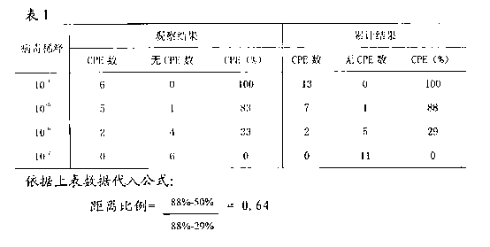
[0019] Detection process: steps of resuscitating and culturing MDCK cells; virus solution dilution, inoculation, red blood cell agglutination; calculation of TCID 50 step;

[0020] (1) Preparation of MDCK cells:

[0021] The sampling density is 2.0×10 6 1.5ml of frozen MDCK cells were placed in a 37°C water bath, shaken to dissolve for 3 minutes, centrifuged at 1000rmp / min for 5 minutes, discarded the supernatant, added 15ml of cell culture medium, and transferred to a 75cm 2 culture flask at 37°C, 5% CO 2 In the incubator, after culturing for 72 hours, the cells were digested with 0.25% EDTA-trypsin, and then diluted with cell nutrient solution (10% fetal bovine serum + DMEM) to 4.0×10 5 The MDCK cell suspension was plated on a 96-well cell plate at a cell density of 40,000 / well (0.1ml / well), and placed at 37°C, 5% CO 2 In the incubator, cultivate for 24 hours to form a dense monolayer...


PUM


Abstract

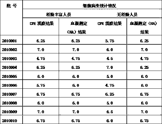
The invention provides a novel determining method of the content of antigen virus of an inactivated vaccine of a recombinant bird flu H5N1 cellgen. The method comprises the following steps of: (1) preparation of MDCK (madin darby canine kidney), wherein 1.5ml of MDCK frozen cell with density of 2.0*10<6> is extracted and put in a water bath at a temperature of 37 DEG C, 10 to 15ml of cell culturefluid is added to culture for 72 hours, the cell is digested with 0.25 percent EDTA (ethylene diamine tetraacetic acid)-pancreatin, the cell nutrient fluid is diluted into MDCK cell suspension of 4.0*10<5>, the MDCK cell suspension is spread to a 96-mesh cell plate in a cell density of 40,000 per pore (0.1ml per pore), the cell plate is put in a CO2 culture case at a temperature of 37 DEG C and humidity of 5 percent to culture for 24 hours to form a compact monolayer for later use; and (2) inoculation of a recombinant bird flu H5N1 antigen, wherein recombinant bird flu H5N1 cellgen antigens in different batches are extracted and respectively added into the compact monolayer with 100mu l in each pore, and 100mu l of cell maintenance fluid is added to culture for four days, and a TCID50 (50% tissue culture infection dose) value is calculated by the number of cytopathic pores in a Reed-Muench method. The method is applicable to a cellgen vaccine for detecting the virus content by an erythrocyte agglutination test, which means that different viruses use corresponding sensitive cells.

Description

technical field [0001] The invention relates to the determination of the virus content of MDCK cells (canine kidney cells) combined with the erythrocyte agglutination test to measure the antigen virus content of the recombinant avian influenza H5N1 cell-derived inactivated vaccine, reducing subjective judgment errors, and has important application value. Background technique [0002] Since the current inactivated vaccine for avian influenza H5N1 is mainly cultivated on chicken embryos, the allantoic fluid is harvested, and the finished vaccine is prepared after inactivation. However, the isolation of influenza virus from chicken embryos or passage can easily cause virus mutation, and the residue of chicken embryos can also cause allergic reactions. Chicken embryos are used as vaccine production substrates, and there are still problems of supply and potential foreign virus contamination during large-scale production. Therefore, Cellular vaccine technology has become an emergi...

Claims


Application Information

Patent Timeline
IPC IPC(8): C12Q1/70C12Q1/02C12R1/93
Inventor 王玉红李延涛袁萍李莉张先锋刘淑梅黄炯潘毅平赵海源
Owner 吉林冠界生物技术有限公司
Why Eureka
Try Eureka