Mycobacterium tuberculosis specific fusion protein as well as preparation and application of mycobacterium tuberculosis specific fusion protein

A Mycobacterium tuberculosis and fusion protein technology, applied in biochemical equipment and methods, hybrid peptides, specific peptides, etc., can solve the problems of low sensitivity and specificity, not exceeding 70%, and achieve high sensitivity, Effect of low cost, high antigen detection sensitivity and specificity

Inactive Publication Date: 2012-10-10
THE 309TH HOSPITAL OF CHINESE PEOPLES LIBERATION ARMY
View PDF2 Cites 5 Cited by
  • Summary
  • Abstract
  • Description
  • Claims
  • Application Information

AI Technical Summary

Problems solved by technology

In addition, other bacterial infections may also produce false positive reactions, resulting in the low sensitivity and specificity of the current commercial tuberculosis antibody detection ki

Method used


Image

  • Mycobacterium tuberculosis specific fusion protein as well as preparation and application of mycobacterium tuberculosis specific fusion protein
  • Mycobacterium tuberculosis specific fusion protein as well as preparation and application of mycobacterium tuberculosis specific fusion protein
  • Mycobacterium tuberculosis specific fusion protein as well as preparation and application of mycobacterium tuberculosis specific fusion protein

Examples


preparation example Construction

[0033] The preparation method of the fusion protein 38kDa-Rv0577-Rv1271c comprises:

[0034] (1) The design of the fusion of three protein epitopes: use molecular biology software to analyze the gene sequence and protein structure of Mycobacterium tuberculosis 38kDa, Rv0577 and Rv1271c, and determine the region, combination and sequence of the three protein epitope fusions; The antigenic epitopes of 38kDa, Rv0577 and Rv1271c proteins were linked in sequence to form a fusion protein. The antigenic epitope of the 38kDa protein is located at the amino terminal of the fusion protein, and the antigenic epitope of the Rv1271c protein is located at the shuttle base of the fusion protein. The DNA sequences of the antigenic epitopes of 38kDa, Rv0577 and Rv1271c in the fusion protein are respectively shown as sequences 1, 2 and 3 in the sequence listing.

[0035] (2) Cloning of fusion of three proteins: Cloning by genetic engineering technology.

[0036] ①Add Xba I restriction site an...

Embodiment 1

[0042] 1. Cloning the 38kDa epitope coding gene by genetic engineering technology:

[0043] 1. Design and synthesize a pair of primers for amplifying the 38kDa epitope according to the sequence 1 in the sequence listing

[0044] Upstream primer (contains restriction endonuclease Xba I site)

[0045] 5'-TGCTCTAGAGGCGGTGGCTCGAAACCACCGAGC-3'

[0046] Downstream primers (containing restriction endonuclease Hind III and Spe I sites)

[0047] 5'-

[0048] CCCAAGCTTCATCATTAACTAGTGCCACCGCTGGAAATCGTCGCGATCAA-3'

[0049] Amplified fragment: 1094bp

[0050] 2. PCR amplification of 38kDa gene

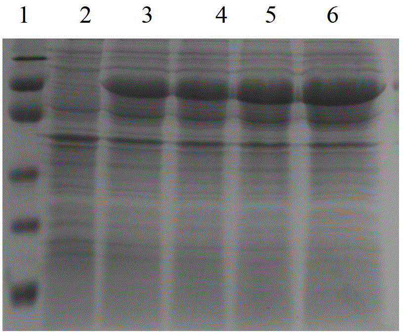
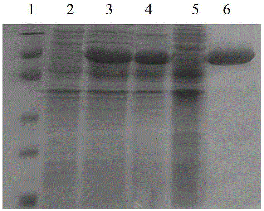
[0051] Using upstream and downstream primers, under the action of Taq plus I DNA polymerase, the 38kDa gene was amplified with Mycobacterium tuberculosis H37Rv genomic DNA as a template. PCR reaction program: 95°C for 5min; 94°C for 1min, 66°C for 1min, 72°C for 2min, cycled 30 times; finally 72°C for 7min. The 1094bp amplified DNA fragment was identified by 1% agarose gel electrophoresis. ...

Embodiment 2

[0170] The 38kDa-Rv0577-Rv1271c fusion protein constructed and purified in Example 1 of the present invention was applied to the serological diagnosis of tuberculosis. It is used as the diagnostic antigen of chemiluminescence immunoassay and applied to the test of clinical specimens, and a good diagnostic effect is obtained. Compared with the three currently commercialized tuberculosis antibody detection kits, it significantly improves the sensitivity of tuberculosis diagnosis. The ELISA detection procedure is as follows:

[0171] 1. Experimental specimens: A total of 103 serum specimens were selected and divided into two groups:

[0172] (1) Tuberculosis group: 54 patients with active tuberculosis diagnosed clinically by imaging, laboratory examination and anti-tuberculosis treatment, including 35 males and 19 females, with an average age of 43.1±18.8 years. Including tuberculosis, tuberculous pleurisy, tuberculous pericarditis, tuberculous meningitis, urinary tuberculosis,...


PUM


Abstract

The invention relates to mycobacterium tuberculosis specific fusion protein as well as preparation and an application of the mycobacterium tuberculosis specific fusion protein and belongs to the technical field of medical immunology diagnosis of tuberculosis. The fusion protein is formed by sequentially connecting antigenic epitopes of three kinds of protein: 38kDa, Rv0577 and Rv1271c, wherein the nucleotide sequence of the antigenic epitope of the protein 38kDa is shown as a sequence 1 in a sequence table, the nucleotide sequence of the antigenic epitope of the protein Rv0577 is shown as a sequence 2 in the sequence table, and the nucleotide sequence of the antigenic epitope of the protein Rv1271c is shown as a sequence 3 in the sequence table. Compared with the commercial antibody detection reagent box in the prior part, the prepared mycobacterium tuberculosis specific fusion protein provided by the invention has the advantages that in the tuberculosis serology diagnosis aspect, the sensitivity is high, the specificity is high, the complementarity with other antigens is realized, and the mycobacterium tuberculosis specific fusion protein can be used for detecting specific antitubercle antibodies in body fluid specimens of serum, hydrothorax and the like.

Description

technical field [0001] The present invention relates to a Mycobacterium tuberculosis-specific fusion protein and its preparation and application, in particular to a Mycobacterium tuberculosis-specific fusion protein 38kDa-Rv0577-Rv1271c prepared by genetic engineering technology and its multi-antigen epitopes. The preparation method belongs to the technical field of tuberculosis medical immunology detection. Background technique [0002] Tuberculosis is still one of the major infectious diseases that endanger human health in the world. Since 1985, the prevalence of AIDS, tuberculosis-infected immigrants and some people living in poverty have caused the incidence of tuberculosis to rise in the United States and other developed countries in Europe and the United States, especially tuberculosis. Bacterial drug resistance and co-infection with human immunodeficiency virus (HIV) make the treatment of tuberculosis even worse. At present, there are about 20 million tuberculosis pa...

Claims


Application Information

Patent Timeline
IPC IPC(8): C07K19/00C12N15/70G01N33/68C12R1/32
Inventor 吴雪琼阳幼荣张俊仙赵卫国冯金栋梁艳
Owner THE 309TH HOSPITAL OF CHINESE PEOPLES LIBERATION ARMY
Why Eureka
Try Eureka