Monoclonal antibody of anti-perkinsus membrane protein, preparation method and application thereof

A monoclonal antibody and membrane protein technology, applied in the field of genetic engineering and immunology, to achieve the effect of a reliable immunological detection method

Inactive Publication Date: 2012-11-07
CHINESE ACAD OF INSPECTION & QUARANTINE
View PDF4 Cites 3 Cited by
  • Summary
  • Abstract
  • Description
  • Claims
  • Application Information

AI Technical Summary

Problems solved by technology

However, there are relatively few reports o

Method used


Image

  • Monoclonal antibody of anti-perkinsus membrane protein, preparation method and application thereof
  • Monoclonal antibody of anti-perkinsus membrane protein, preparation method and application thereof
  • Monoclonal antibody of anti-perkinsus membrane protein, preparation method and application thereof

Examples


Embodiment 1

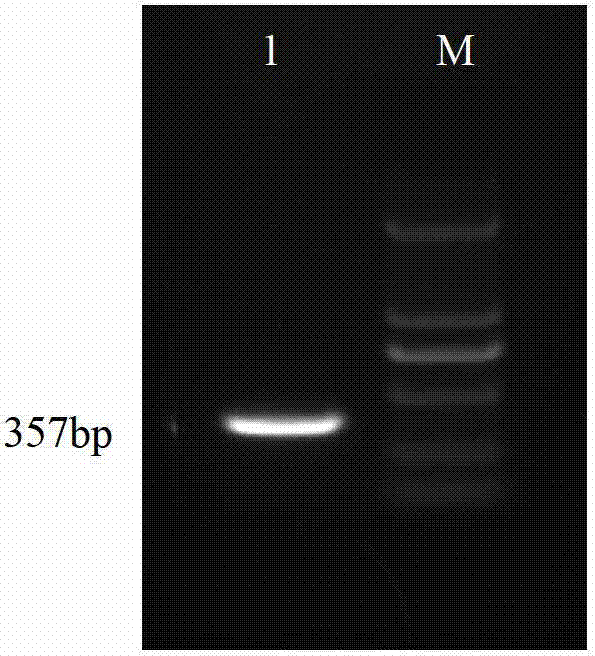
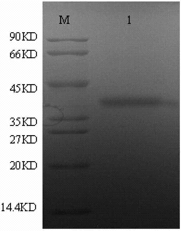
[0027] Example 1 Obtaining of Peyrin's membrane protein (MOE)

[0028] 1.1 Primer design

[0029] According to the sequence published in ACCESSION No.EF632302 in the NCBI Gene Bank (GenBank) of the National Center for Bioinformatics in the United States, primers were designed at both ends of the open reading frame. The primer sequence is:

[0030] CB5D4UP: 5'-GGCTGATATC GGATCC TCCTCTTGTCCCACAGGGGATGC-3' (the underline is the BamHI restriction site)

[0031] CB5D4LP: 5'-CCGCAAGCTT GTC GAC TTATGACGTAGGACATGTCGG-3' (underlined is the SalI restriction site)

[0032] 1.2 Preparation of primers

[0033] After artificially synthesizing the primers, the primers were diluted to 10 μmol / l, and the upstream and downstream primers were mixed in equal volumes at a ratio of 1:1, and stored at -20°C after aliquoting.

[0034] 1.3 Extraction of the DNA of Piedia worms

[0035] Select unhealthy or just dead Philippine clams infected with Pichinella, separate gill tissues, and extract D...

Embodiment 2

[0045] Example 2 Obtaining of Monoclonal Antibody Against Pichinensis membrane protein (MOE)

[0046] 1.1 Immunization of mice with purified Pietchenia membrane protein (MOE)

[0047] Mix the purified membranous membrane protein (MOE) and complete Freund's adjuvant in equal volumes, oscillate, emulsify, and grind into a water-in-oil chylus. to the water-in-oil state).

[0048] 1) Initial immunization: 1-80 μg of specific Pijenia membrane protein (MOE) plus Freund's complete adjuvant subcutaneously injected into mice at multiple points (generally 0.25-1 mL, 0.2 mL / spot);

[0049] 2) Second immunization: 1 week later, the dose was halved, and the mice were subcutaneously injected with Freund's incomplete adjuvant in multiple points;

[0050] 3) The third immunization: 3 days later, the dose was the same as above, and the mice were subcutaneously injected with incomplete Freund's adjuvant at multiple points (5-7 days later, blood was collected to measure the potency and detect ...

Embodiment 3

[0107] Example 3 Determination of the titer of the monoclonal antibody against Pichinensis membrane protein (MOE) (ELISA detection method)

[0108] 1) Coating: add 2 μg / ml antigen (namely the specific Pichenya membrane protein (MOE) prepared in Example 1), 100 μl / well, overnight at 4°C, wash with washing solution 3 times.

[0109] 2) Blocking: add 150 μl / well blocking solution, wash 3 times after 2 hours at 37°C, and pat dry. Store in a 4°C refrigerator for later use.

[0110] 3) Add the sample to be tested:

[0111] a. For the detection of serum / antibody / ascitic fluid titer, the first well was diluted 1:1000, followed by 1:3 gradient dilution, incubated at 37°C for 30min, washed 4 times, and patted dry.

[0112] b. For the detection of cell supernatant, take 100 μl of cell supernatant, add it to the corresponding microplate, incubate at 37°C for 30 min, wash the plate 4 times, and pat dry.

[0113] 4) Add secondary antibody: take horseradish-enzyme-labeled goat anti-mous...


PUM


Abstract

The invention provides a monoclonal antibody of anti-perkinsus membrane protein (MOE). The monoclonal antibody of the anti-perkinsus membrane protein (MOE) is secreted by a hybridoma cell strain IAQ1101 of the anti-perkinsus membrane protein (MOE) with an accession number of CGMCC (China General Microbiological Culture Collection) No.6180. The invention also provides a preparation method and application of the monoclonal antibody. The monoclonal antibody of the anti-perkinsus membrane protein (MOE) is obtained by a genetic engineering technology and an immunological method; by labeling the monoclonal antibody with fluorescence, the immunofluorescence labeling technology is utilized to quickly and accurately detect infected perkinsus in shellfish, accordingly, a reliable immunological detection method is provided for the rapid identification of a port.

Description

technical field [0001] The present invention relates to the fields of genetic engineering and immunology, in particular, to a monoclonal antibody against Pichinensis membrane protein (MOE), its preparation method and application. Background technique [0002] Perkinsus (or clapper worm) is one of the main pathogens affecting the development of shellfish farming in the world. Due to its wide distribution and serious harm, it is stipulated by the World Organization for Animal Health (OIE) as a must-report aquatic animal disease. In 1997, Pijtella was detected in clams imported from the Philippines in my country. In 2001, Liang Yubo et al. used thioglycollate medium (FTM) to culture and tissue slices to prove that the infection rate of clams was 20%-100%. In 2008, for the first time, the Ministry of Agriculture listed Pichet worms as the parasitic pathogens that mainly infect clams. [0003] In view of the seriousness of Piedia worm damage, a lot of research work has been car...

Claims


Application Information

Patent Timeline
IPC IPC(8): C07K16/20G01N33/577G01N33/569C12N5/20
Inventor 王彩霞吴绍强林祥梅景宏丽张永宁吕继洲邓俊花
Owner CHINESE ACAD OF INSPECTION & QUARANTINE
Why Eureka
Try Eureka