
Method and device for detecting tumorous tissue in the gastrointestinal tract with the aid of an endocapsule

A technology of inner capsule and gastrointestinal tract, applied in the direction of character and pattern recognition, endoscope, radio detector in the body, etc.

Active Publication Date: 2013-02-27
SIEMENS HEALTHCARE GMBH +1
View PDF9 Cites 0 Cited by
  • Summary
  • Abstract
  • Description
  • Claims
  • Application Information

AI Technical Summary

Problems solved by technology

This different factor and biomolecular cell structure has a strong influence, and the correlation of the cellular organization as healthy or neoplastic diseased cellular organization cannot be achieved with sufficient reliability.

Method used


Image

  • Method and device for detecting tumorous tissue in the gastrointestinal tract with the aid of an endocapsule
  • Method and device for detecting tumorous tissue in the gastrointestinal tract with the aid of an endocapsule
  • Method and device for detecting tumorous tissue in the gastrointestinal tract with the aid of an endocapsule

Examples


Embodiment Construction

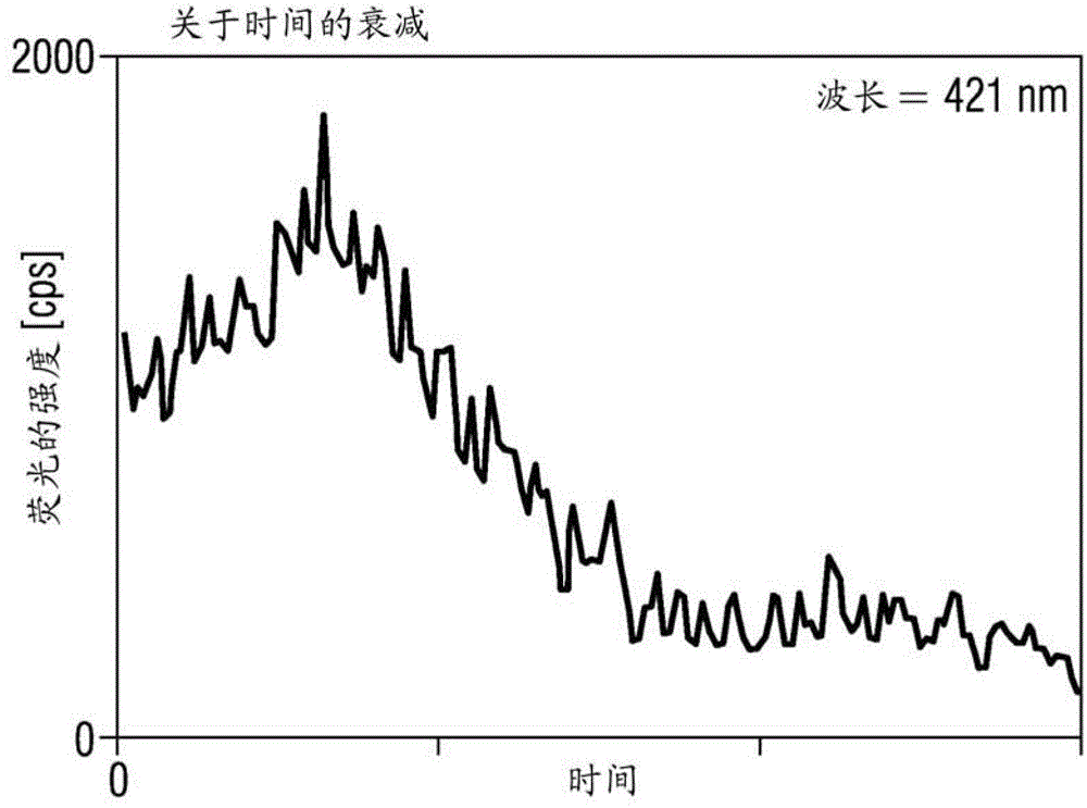
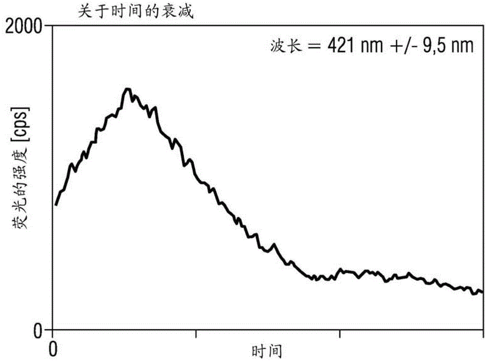
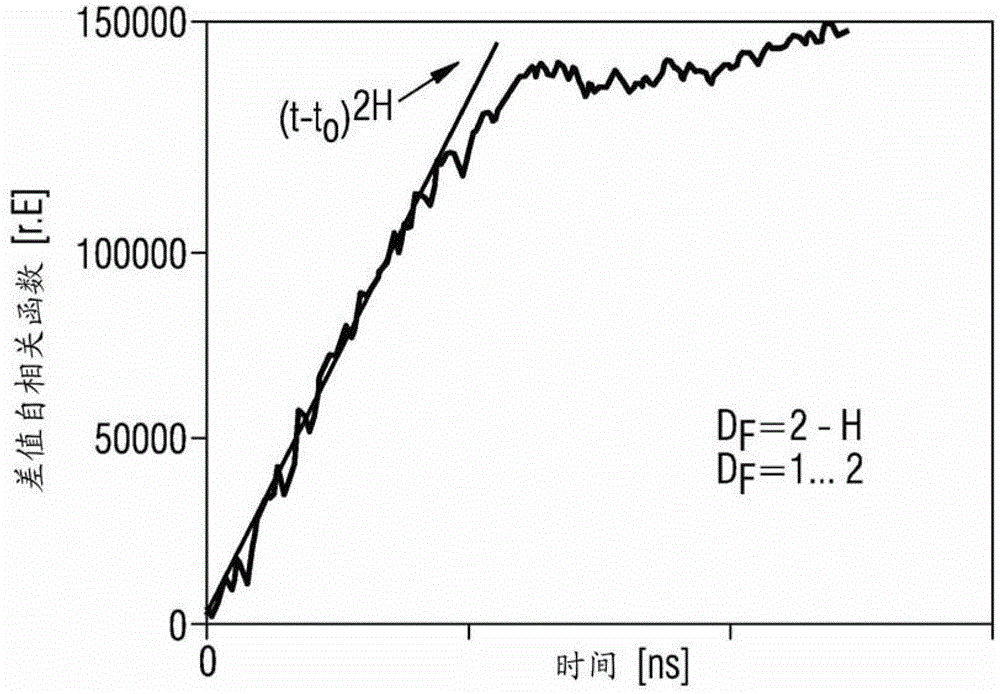
[0044] according to Figure 1 to Figure 3 The graph for is based on examinations performed in vitro for reasons of simplification and not in vivo for reasons of reproducibility. The cell tissue sample is placed in a tank serving as a cell tissue sample container, and the electromagnetic radiation is directed to a predetermined position of the cell tissue sample by means of an optical fiber. As radiation source a nitrogen laser is used. The wavelength of electromagnetic radiation used for autofluorescence excitation of tissue is 337 nm.

[0045] The harvested cell samples were cooled to a temperature of 15°C in order to delay necrosis and were kept at this temperature at least until the end of the examination.

[0046] After the radiation source is turned off at t 0 At this point, the electromagnetic radiation emitted as a result of the autofluorescence of the cellular tissue is directed on the same optical fiber to a spectrometer with which detection in the wavelength inter...


PUM

PropertyMeasurementUnit
Wavelengthaaaaaaaaaa

Abstract

The invention relates to a method for detecting tumorous cell tissue in the gastrointestinal tract, in which method electromagnetic radiation is emitted in a locally defined manner onto cell tissue with the aid of an endocapsule, and, after the radiation source has been switched off, the decay behaviour of the cell tissue's inherent fluorescence intensity, excited by the electromagnetic radiation, is detected on the cell tissue by a detector, with temporal and spectral resolution and with a known scanning rate or known scanning rates for at least one wavelength, and, with the intensity measurement values that are obtained, the difference autocorrelation function C(t) of the intensity decay behaviour is determined, from which the fractal dimension DF for the respective irradiated cell tissue is determined, and the value of the fractal dimension DF is used for a classification in respect of a tumorous invasion of the respective irradiated cell tissue. The invention further relates to a device comprising an endocapsule with which electromagnetic radiation from a radiation source can be emitted onto cell tissue of the gastrointestinal tract.

Description

technical field [0001] The present invention relates to a method for identifying the cellular tissue of a neoplastic lesion in the gastrointestinal tract by means of an inner capsule. Background technique [0002] To identify cancer in the gastrointestinal tract, a tissue sample is taken, for example during a gastroscopy, and tested for the presence of cancer. Here, multiple biopsies are frequently requested. To reduce this frequency, so-called autofluorescent inner capsules are used, in which the body's own fluorescence phenomenon due to the high concentration of high mass exchange activity in the malignant tissue is used. A further possibility for biopsy control is the use of an endoscope, ie examination by means of a microscope integrated in the endoscope, in which the patient has to be given a contrast agent for staining the tissue. But in both cases a biopsy is required. [0003] The obtained tissue samples are examined histologically in the laboratory. In this case...

Claims


Application Information

Patent Timeline
IPC IPC(8): A61B5/00A61B5/07G06K9/00
CPCA61B1/043A61B1/041G06K2209/053G06K9/00147A61B5/0071G06T7/00G01N21/6486A61B5/4205A61B5/0084G01N21/6408A61B5/0075G06V20/698G06V2201/032A61B5/0059
Inventor J.费尔R.南科
Owner SIEMENS HEALTHCARE GMBH
Learn More