Degradation of PA200 and acetylation-mediated core histones through proteasome

An acetylation and histone technology, applied in the fields of peptide/protein components, hydrolase, drug combination, etc., can solve the problems of unclear acetylation spermatogenesis and DNA repair

Active Publication Date: 2013-07-10
BEIJING NORMAL UNIVERSITY
View PDF5 Cites 8 Cited by
  • Summary
  • Abstract
  • Description
  • Claims
  • Application Information

AI Technical Summary

Problems solved by technology

Although histone acetylation sites and acetylases or sirtuins have been extensively studied, how acetylation regulates transcription, spermatogenesis, and DNA repair remains unclear

Method used


Image

  • Degradation of PA200 and acetylation-mediated core histones through proteasome
  • Degradation of PA200 and acetylation-mediated core histones through proteasome
  • Degradation of PA200 and acetylation-mediated core histones through proteasome

Examples


Embodiment 1

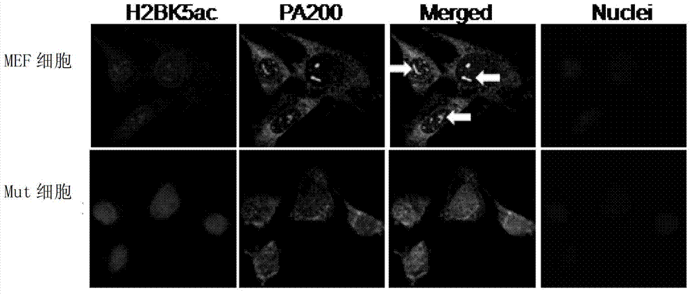
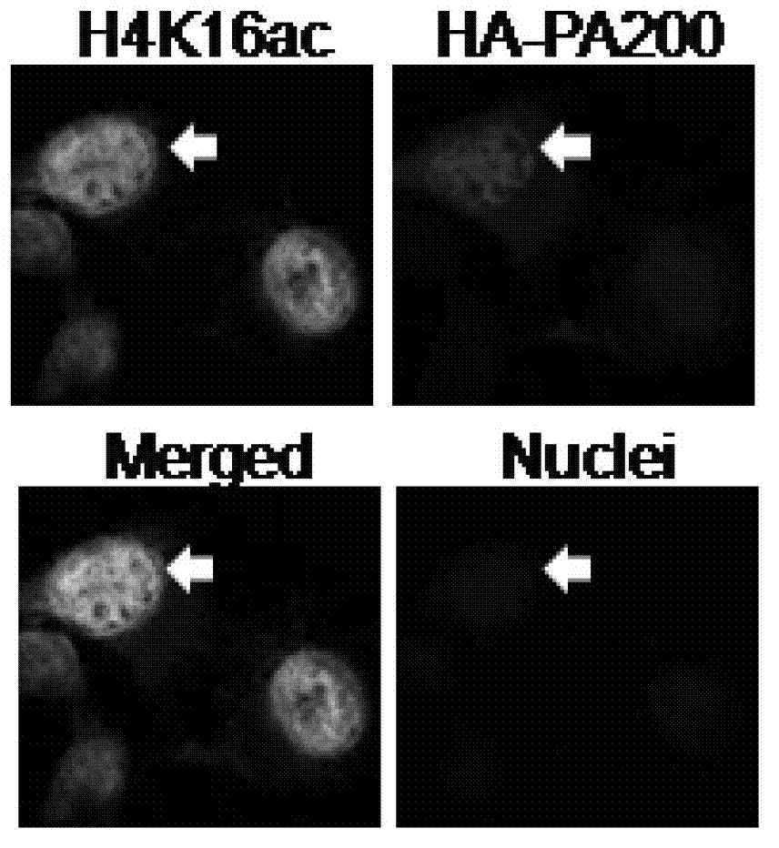
[0063] Example 1. PA200 recognizes and binds to acetylated histones

[0064] 1. Co-localization of PA200 and H2BK5ac

[0065] Preparation method of mouse H2BK5ac antibody: H2BK5ac synthesized by GenScript (Nanjing, China) (H2BK5ac represents histone H2B acetylated at the K5 site, with a biotin label, specifically: PEPAKacSAPAPKKGSKKAVTKA-Biotin) immunized BALB / C Mice, collect serum.

[0066] Take MEF cells or Mut cells and use mouse H2BK5ac antibody (the corresponding secondary antibody is goat anti-mouse-Dylight594, red) and rabbit PA200 antibody (the corresponding secondary antibody is goat anti-rabbit-FITC, green) for immunofluorescence Stain, DAPI stains the nucleus.

[0067] See you in the photo figure 1 (The arrow indicates a trajectory for each cell). In MEF cells, PA200 antibody recognizes punctate structures in the nucleus, and H2BK5ac also appears in this area. The staining intensity of H2BK5ac in Mut cells was significantly greater than that of MEF cells. The results sh...

Embodiment 2

[0112] Example 2. Identification of two different types of proteasomes in mammalian testes

[0113] Histones are lost in large amounts in spermatogenic cells. In order to find the potential mechanism of histone degradation, this example detects whether mammalian spermatogenic cells have a unique proteasome form.

[0114] 1. Extract the total protein of bovine skeletal muscle (muscle), which is total muscle protein; extract the total protein of bovine testicular seminiferous tubules, which is total spermatogenic protein.

[0115] 2. Perform non-denaturing gel electrophoresis on total muscle protein and total spermatogenic protein, and incubate with fluorescent peptide substrate (succinyl LLVY-7-amino-4-methylcoumarin) (for co-incubation, set two treatments, one Add SDS with a final concentration of 0.02% during the incubation, and do not add SDS during the other incubation; after shaking at room temperature for 15 minutes, observe the proteasome band under UV light.

[0116] See the re...

Embodiment 3

[0132] Example 3. Specific subunits and activities in the spermatogenic proteasome

[0133] 1. C2C12 myoblasts, TM3 Leydig cells, TM4 Sertoli cells, mouse GC-1spg type B spermatogonia (GC1 cells for short), mouse GC-2spd(ts) spermatocytes (GC2 cells for short) ), sperm (sperm), testis tissue (testis), spleen tissue (spleen), respectively extract total protein and perform SDS-PAGE electrophoresis and immunoblotting analysis. Each of the above cells were purchased from the American Model Culture Collection Bank. All the above tissues are the tissues of BALB / C mice. The primary antibodies used in western blotting were rat anti-α4s antibody, rat anti-α4 antibody, β1i antibody, β1 antibody, β2i antibody, β2 antibody, β5i antibody, β5 antibody, PA28α antibody, PA200 antibody, Rpt4 antibody, Rpn7 antibody and GAPDH antibody.

[0134] See the result Figure 8 A. α4s and β2i can only be detected in Sertoli cells and spermatocytes, while β5i, PA200, 19S subunits (such as Rpt2, Rpt4 and R...


PUM


Abstract

The invention discloses an application of PA200 proteins in preparation of products. The products have at least one of the following functions from (1) to (4): (1) combining with acetylated proteins; (2) promoting the degradation of the acetylated proteins; (3) participating in repair of DNA (deoxyribonucleic acid) damages of somatic cells; and (4) participating in the formation of sperms. The invention has breakthroughs in four following aspects: (1) finding a mechanism of regulation and control of degradation of the histones, the sperm formation and the DNA repair by acetylation; (2) revealing the degradation of the histones medicated by acetylation rather than ubiquitination through PA200 / Blm10 proteasome; (3) finding novel testis-specific proteasome containing PA200 and alpha 4s, and revealing the core histones as a first type biological substrate for the proteasome; and (4) revealing that a histone deacetylase inhibitor can promote the degradation of the histones which are induced by DNA double-strand breaks and medicated by acetylation, enhance the sensitivity of cells to DNA damages and easily lead to the death of the cells.

Description

Technical field [0001] The present invention relates to PA200 and acetylation-mediated degradation of core histones by the proteasome. Background technique [0002] The proteasome catalyzes the ATP and polyubiquitination-dependent degradation of most proteins in cells, and also catalyzes the production of histocompatibility complex class I antigen peptides in vertebrates. The 26S proteasome is composed of two subcomplexes, namely the 20S catalytic particle and the regulating particle that binds one or both ends of it. There are two main forms of 20S core particles that have been identified in mammals: constitutive proteasome and inducible immune proteasome. The former contains three catalytic subunits (β1, β2 and β5), while the latter contains three subunits (β1i, β2i and β5i) closely related to induction. The main regulatory particle of the proteasome is the 19S complex, which can bind to the polyubiquitinated protein, unfold it and transfer it into the 20S catalytic particle....

Claims


Application Information

Patent Timeline
Patent Type & Authority Applications(China)
IPC IPC(8): A61K38/16A61K38/48C07K14/00C12N9/50A61K45/00A61K45/06A61P15/16A61P35/00A61P15/08
CPCA61K38/17A61P15/08A61P15/16A61P35/00C07K14/47
Inventor 邱小波钱民先逄也刘翠华王广菲朱倩倩张晓旭刘珊
Owner BEIJING NORMAL UNIVERSITY
Why Eureka
Try Eureka