Novel ultrahigh resolution photoelectric integration micro-imaging system

A technology of photoelectric fusion and microscopic imaging, applied in microscopes, optics, optical components, etc.

Active Publication Date: 2013-09-18
INSITUTE OF BIOPHYSICS CHINESE ACADEMY OF SCIENCES
View PDF8 Cites 24 Cited by
  • Summary
  • Abstract
  • Description
  • Claims
  • Application Information

AI Technical Summary

Problems solved by technology

[0008] 2. Unable to continuously perform high-precision fluore

Method used


Image

  • Novel ultrahigh resolution photoelectric integration micro-imaging system
  • Novel ultrahigh resolution photoelectric integration micro-imaging system
  • Novel ultrahigh resolution photoelectric integration micro-imaging system

Examples


Embodiment Construction

[0057] In order to make the objectives, system solutions and advantages of the present invention more clearly understood, the present invention will be further described in detail below in conjunction with specific embodiments and with reference to the accompanying drawings.

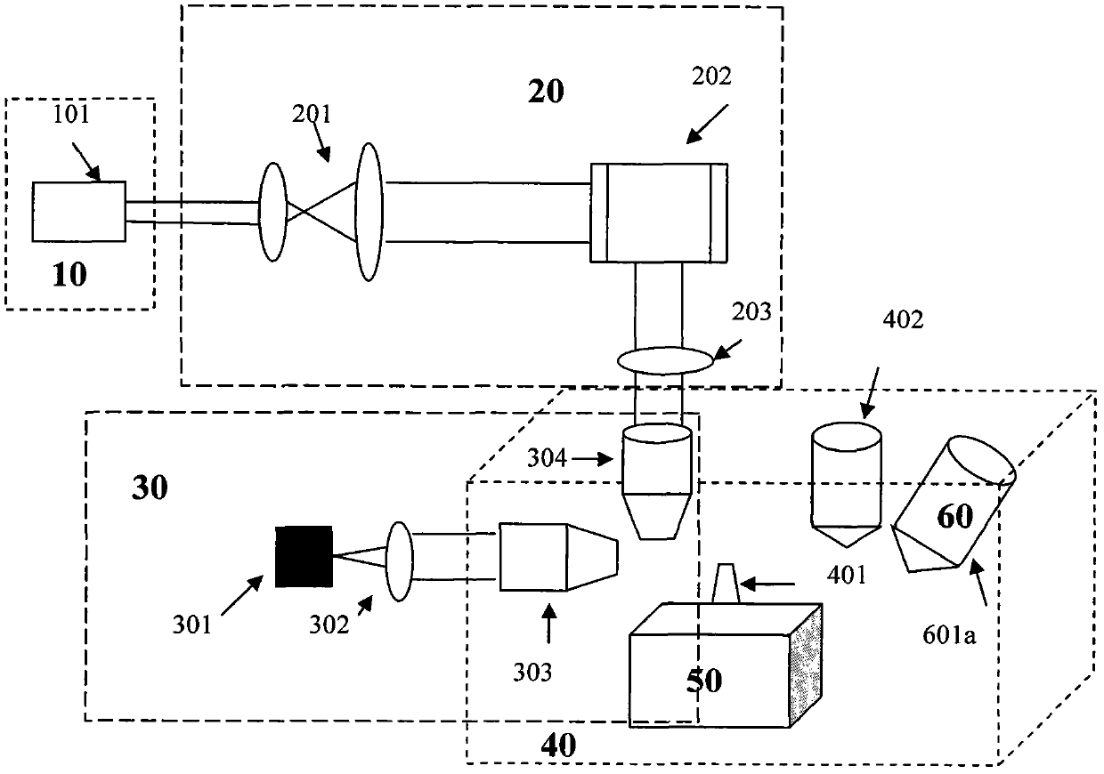
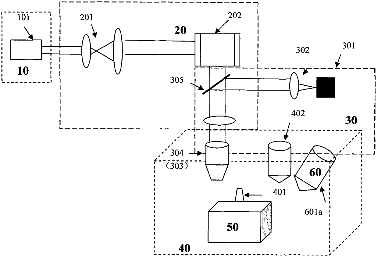
[0058] in the present invention figure 1 In an exemplary embodiment, a schematic structural diagram of a photoelectric fusion microscope imaging system in which an illumination objective lens and an imaging objective lens are different objective lenses is provided (eg, a slice light scanning microscope). The photoelectric fusion microscope imaging system includes: a fluorescence microscope system, a sample transfer system, a sample slice system and a scanning electron microscope system. The fluorescence microscope system includes a light source system 10 , a scanning beam expander system 20 , and a fluorescence imaging system 30 . Wherein: the scanning beam expander system 20 expands the laser beam and s...


PUM


Abstract

The invention discloses a photoelectric integration micro-imaging system combined by a high-resolution fluorescence microscope and a scanning electron microscope. The photoelectric integration micro-imaging system comprises a fluorescence microscope system, a sample transfer system, a sample cutting system and a scanning electron microscope system, wherein the fluorescence microscope system comprises a light source system, a scanning and beam-expanding system and a fluorescence imaging system; the sample transfer system is used for transferring samples between the fluorescence microscope system and the scanning electron microscope system; the sample cutting system is used for performing uniform and ultrathin cutting on the samples; the scanning electron microscope system is used for acquiring electron scanning images of the samples. According to the system, sample structure information and biomolecule positioning information are fused, and the samples are cut to be ultrathin by utilizing the cutting system, so that the samples can be imaged in the electron microscope and the fluorescence microscope system, and three-dimensional image information of the samples is acquired. The light-sensitive positioning fluorescence imaging, ultrathin cutting and other ultrahigh-resolution fluorescence microscope technologies and the scanning electron microscope technology are organically combined, and the multiple fluorescence microscope-scanning electron microscope integration imaging systems can be formed.

Description

[0001] System field: [0002] The invention relates to the technical field of optical microscopic imaging and electron microscopic imaging, in particular to a photoelectric fusion microscopic imaging system using a combination of a fluorescence microscope system and a scanning electron microscope. Background technique: [0003] Electron microscopy is an important research method for structural biology research, enabling biologists to study the ultrafine structure of organisms and their relationship with function at the nm scale, but it is impossible to clarify the types and distribution of biomolecules in different ultrastructures—— Although immunocolloidal gold technology can label target molecules on the ultrastructure, its wide application is limited due to its low labeling efficiency, low resolution, and nonspecific labeling. [0004] Fluorescence imaging is an important research method in modern life science research, which can obtain the positioning information, function...

Claims


Application Information

Patent Timeline
IPC IPC(8): G01N21/64G02B21/36G02B21/06
Inventor 徐涛罗志勇纪伟仓怀兴孟涛张翔季刚付彦辉贾策
Owner INSITUTE OF BIOPHYSICS CHINESE ACADEMY OF SCIENCES
Why Eureka
Try Eureka