Micro-elasticity imaging method based on tissue microbubble dynamics model

A dynamic model, elastography technology, applied in the field of biomedical ultrasound imaging, can solve the problems of inconsistency of actual elastic distribution, strain estimation error, inability to distinguish small-sized thin tissue in the body, etc., so as to improve the contrast tissue ratio. , The effect of inhibiting tissue signal and high detection sensitivity

Active Publication Date: 2013-10-02
XI AN JIAOTONG UNIV
View PDF4 Cites 11 Cited by
  • Summary
  • Abstract
  • Description
  • Claims
  • Application Information

AI Technical Summary

Problems solved by technology

The limitations of existing elastography methods mainly include: (1) In static compression elastography, the internal deformation of the tissue is mainly determined by the boundary conditions of the external force and the actual elastic distribution. The distributions do not match, leading to significant strain estimation errors
(2) In dynamic compression elastography, a small-scale elastic distribution can be calculated by locally applying excitation, but the imaging resolution will be limited by the size of the excitation source and the imaging speed
(3) The resolution of the existing elastography technology is basically at the millimeter level, which is temporarily unable to meet the needs of distinguishing the elastic changes of small thin-layer tissues in the body, such as blood vessel walls, epithelial tissues, and gastric mucosa.

Method used


Image

  • Micro-elasticity imaging method based on tissue microbubble dynamics model
  • Micro-elasticity imaging method based on tissue microbubble dynamics model
  • Micro-elasticity imaging method based on tissue microbubble dynamics model

Examples


Embodiment Construction

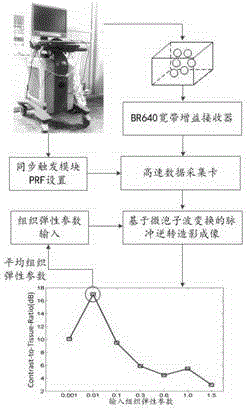
[0028] refer to figure 1 , a microelastography method based on a microbubble dynamics model in tissue, comprising the steps of:

[0029] (1) Connect the linear array transducer set on the detection target with the main control computer through a programmable ultrasonic device with a radio frequency data acquisition interface;

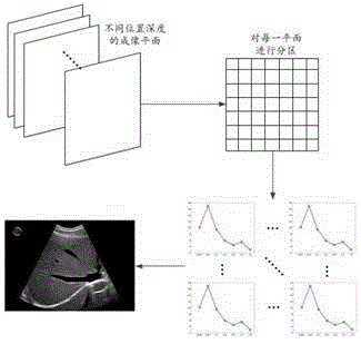
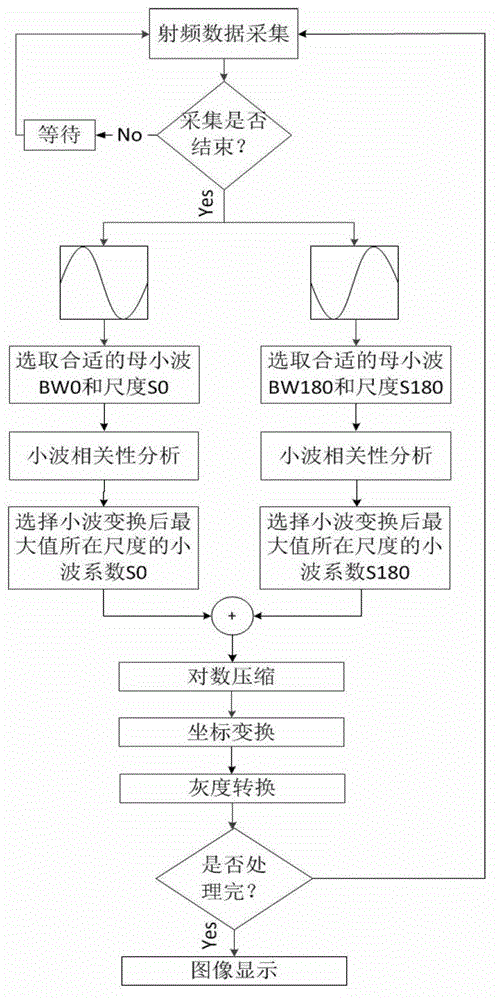
[0030] (2) Select a microbubble dynamics model that can reflect tissue elasticity parameters, test the model through experimental means, and conduct sensitivity analysis and reliability evaluation on it, and finally select a model that is sensitive to tissue elasticity parameters and has high reliability Construct the mother wavelet and solve it to obtain the change curve of the microbubble vibration radius with time and the radial vibration velocity and acceleration, and calculate the sound pressure radiated by the vibrating microbubble according to the volume change rate with time: Where P is the sound pressure radiated by the vibrating microbubble,...


PUM


Abstract

The invention provides a micro-elasticity imaging method based on a tissue microbubble dynamics model. According to the invention, a mother wavelet which has strong correlation with a microbubble signal and weak correlation with a tissue signal is established according to that microbubble vibration is influenced by characteristics of a surrounding tissue, and a tissue microbubble dynamics model can be used for establishing the relation of a microbubble vibration signal and tissue elasticity, the microbubble signal is detected for imaging through a pulse inversion and wavelet transform combined imaging algorithm, and when the detection signal is the closest to a model signal, the maximum tissue contrast ratio can be obtained, so that the elasticity parameters of the tissue within the range of a dozen to tens of microns around the tissue are obtained in a reverse derivation manner. The method can be applied to real-time monitoring on a high-intensity focused ultrasound therapeutic process and elasticity detection of a biological thin-layer tissue, can overcome the limitations that the general elasticity imaging requires external pressure, and is influenced by boundary conditions easily, and effectively improves the imaging resolution to the micron level from the millimeter level.

Description

technical field [0001] The invention belongs to the technical field of biomedical ultrasonic imaging, and in particular relates to a microbubble vibration signal detection based on a microbubble dynamics model in tissue, a system and a method for reverse calculation of tissue parameters around the microbubble and microelastic imaging. Background technique [0002] Elastography is mainly based on the difference in the elastic coefficient of different tissues and the degree of tissue strain after being subjected to stress to display and locate the elastic changes caused by lesions. It is of great value to monitor tissue solidification caused by thermal ablation, such as high-intensity focused ultrasound. Ultrasound elastography is currently the most important elastography method, and nuclear magnetic resonance can also be used for elastography. Ultrasound elastography mainly includes: (1) static / quasi-static compression elastography, (2) acoustic radiation force dynamic compr...

Claims


Application Information

Patent Timeline
Patent Type & Authority Applications(China)
IPC IPC(8): A61B8/08
Inventor 万明习乔阳紫
Owner XI AN JIAOTONG UNIV
Why Eureka
Try Eureka