Preparation method and application of kit for double-sandwich immunofluorescence quantitative detection of human anti-Mullerian hormone (AMH) on basis of quantum dots
An immunofluorescence and quantitative detection technology, which is applied in the field of immunodiagnosis, can solve the problems of ineffective clinical promotion, many influencing factors, and not widely deployed, and achieve effective detection and risk assessment, improve resolution, and provide the effect of resolution
Inactive Publication Date: 2014-04-16
李志荣
3 Cites 17 Cited by
AI-Extracted Technical Summary
Problems solved by technology
Some domestic diagnosis and treatment institutions have introduced the kit and conducted relevant clinical tests, but its price is relatively expensive and cannot be effectively promoted clinically
At present, there is no registered kit supply using quantum dot immunoassay method to detect AMH in China, and t...
Method used
Detecting result is shown in the above table, and the minimum detection amount obtained by substituting the result into standard curve equation calculation by the above method is 0.575pg/ml, showing that kit of the present invention and detection method have high sensitivity to the detection of people's AMH.
[0032] Add appropriate amount of lysate in the dry precipitation of the bacterium, and ensure that the protein is always in the ice bath during the ultrasonic cracking thalline ultrasonic process under the ice bath, so as to prevent the overheating of the ultrasonic wave from affecting the stability of the protein. Centrifuge at 12000rpm at 4°C to collect supernatant protein, identify protein and detect protein expression by Western Blot.
[0039] Two adult rabbits were prepared, and 100 μg of purified recombinant human AMH antigen was dissolved in 1 ml of phosphate buffer solution for use. Add mycobacteria to 1ml of Freund's incomplete adjuvant to make a complete adjuvant, add 1ml of antigen solution, shake vigorously to make it fully emulsified, draw out the emulsion with a 3ml syringe, connect it with a 25G need...
Abstract
The invention discloses a kit for double-sandwich immunofluorescence quantitative detection of human anti-Mullerian hormone (AMH) on the basis of quantum dots and application thereof. The original kit contains an AMH protein standard substance, an ELISA plate coated with an AMH specific polyclonal antibody and a CdTe quantum dot labeled AMH monoclonal antibody. The detection antibody used in the kit is a CdTe quantum dot label for monoclonal antibodies, and can better remove background signals; the detection method is simple and convenient, and has high practicality; the detection result directly determined by a fluorescent ELIASA indicates that the emitted fluorescence has the advantages of narrow spectrum peak, weak autofluorescence and high sensitivity; the kit has the advantages of high fluorescence intensity and long stabilization time, and overcomes the actual states of low sensitivity and poor specificity in the traditional ELISA detection kit; and the kit can enhance the resolution, sensitivity and specificity of ovary reservation function detection.
Application Domain
Disease diagnosisBiological testing +1
Technology Topic
Polyclonal antibodiesAnti-Müllerian hormone +13
Image
Examples
- Experimental program(1)
Example Embodiment
[0021] The present invention will be further described below in conjunction with embodiments, but it is not limited thereto.
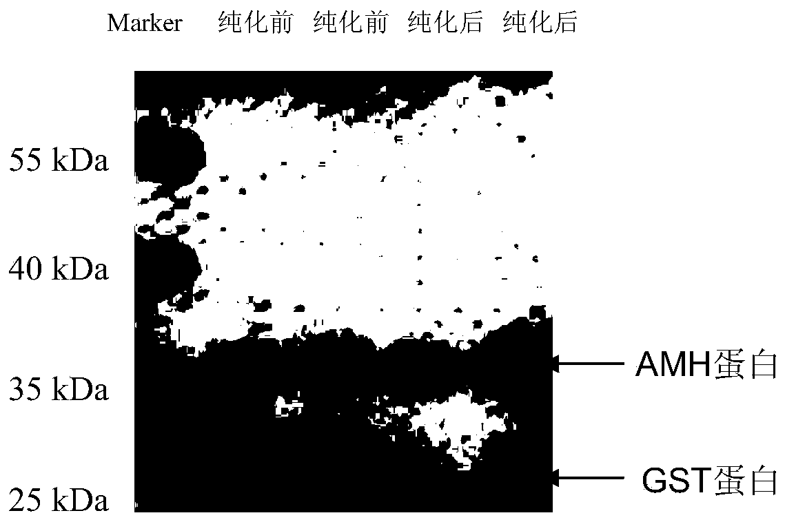
[0022] 1) Construction of AMH prokaryotic expression vector
[0023] The immunogenic region of AMH was selected. The gene sequence is shown in SEQ ID NO. 2, with a total of 297 bp, and the corresponding amino acid sequence is shown in SEQ ID NO. 1, with a total of 93 amino acids.
[0024] The specific primers designed to amplify the sequence of the AMH immunogen region are as follows:
[0025] EcoR I Up primer: CCG GAATTC AGCGTAGACCTCCGCGCCGC (the underlined part is the recognition sequence of the endonuclease EcoR I) (SEQ ID NO.3),
[0026] Xho I Down primer: CCG CTCGAG CCGGCAGCCACACTCGGTGG (the underlined part is the recognition sequence of endonuclease Xho I) (SEQ ID NO. 4).
[0027] The total RNA of human ovarian cancer cells was extracted by chloroform, and the AMH immunogenic sequence was amplified by reverse transcription and PCR primers. The amplified AMH sequence was digested and ligated to the pGEX-4T-1 prokaryotic expression vector to transform it into Among the DH5α competent cells, the transformed cells were inoculated into ampicillin-resistant LB solid medium with a spreading rod, and cultured overnight at 37°C. Monoclonal colonies were picked and a small amount of amplification was carried out. The plasmids were extracted, and EcoRI and XhoⅠ were used. After digesting the plasmid, it was verified by agarose electrophoresis that if a band of about 300 bp can be cut out, it will be initially identified as a colony containing the target gene, and further sequenced to verify that the sequence is correct, that is, the prokaryotic recombinant fusion protein expression vector of AMH has been successfully obtained. It is pGEX-4T-1-AMH.
[0028] 2) Expression and purification of AMH immunogen
[0029] Expression of AMH target protein
[0030] Transform pGEX-4T-1-AMH into Escherichia coli RG2, pick the clones at 37°C, shake culture at 250rpm until the OD value reaches 0.4~0.6, incubate at 10°C and 150rpm under 0.5mM IPTG induction overnight, and then centrifuge at 5000g for 10min to collect the bacteria spare. Dry bacterial pellets can be stored at -80°C.
[0031] AMH target protein expression detection
[0032] Add an appropriate amount of lysis solution to the dry bacterial pellet, and ensure that the protein is always in the ice bath during the ultrasonic lysis of the bacterial body under the ice bath to prevent the overheating of the ultrasound from affecting the protein stability. Centrifuge at 12000 rpm and 4°C to collect the supernatant protein. Western Blot identifies the protein and detects the protein expression.
[0033] Separation and purification of AMH target protein
[0034] Add an appropriate amount of lysis solution to the dry bacterial pellet, and ultrasonically lyse the bacteria under ice bath to ensure protein during the ultrasonic process
[0035] Stay in an ice bath to prevent overheating of ultrasound from affecting protein stability. Centrifuge at 12000rpm, 4℃, collect the supernatant protein, and filter at 0.44um. Mix the protein lysate collected by centrifugation and the pre-washed GSH-agarose thoroughly, and incubate for 4 hours with shaking at 4°C. The cell lysate was washed 3 times, TBS washed 3 times, and 10mM glutathione eluate was added to elute the protein. The eluted protein can pass through a 10KD protein concentration column to concentrate the protein and remove the glutathione in the liquid, and wash with PBS. The purified protein was measured with Coomassie Brilliant Blue method and its purity was identified by western blot (see figure 1 ).
[0036] 2. Preparation and identification of rabbit anti-human AMH polyclonal antibody
[0037] 1) Preparation of rabbit anti-human AMH polyclonal antibody
[0038] Animal immunity
[0039] Prepare two adult rabbits, and dissolve 100 μg of purified recombinant human AMH antigen into 1 ml phosphate buffer solution for later use. Add mycobacteria to 1ml Freund’s incomplete adjuvant to make a complete adjuvant, and add 1ml of antigen solution, shake vigorously to make it fully emulsified, use a 3ml syringe to draw the emulsion, connect a 25G needle, and eliminate air bubbles in the syringe . Take the rabbit out of the cage and place it on a flat place. Inject subcutaneously in 4 different locations, two on the back and two on the thigh. The rabbit hair at the injection site was stroked and the exposed skin was disinfected with ethanol. Pinch out the skin, insert the needle into the needle at an angle of 15 degrees relative to the skin. The depth of the needle is 1 cm to 2 cm. Be careful not to penetrate the muscle. Inject about 500 μl of antigen solution into 4 different parts. After the injection, place the needle at the injection site for a few seconds and then gently pull it out, and disinfect the injection site with ethanol. Repeat the above operation in 4 places. Antigen was injected every 4 to 6 weeks, and blood was collected 7 to 10 days after injection. Compare the blood collected with the blood collected before the injection to check for antibody production. A large amount of blood can be collected after it is determined that antibodies are produced, but each rabbit should not collect more than 40ml of blood to prevent shock.
[0040] Collect serum
[0041] Gently put the rabbit into the holder, apply xylene to the upper and middle part of the ear blood vessel, and cut a 0.23cm~0.3cm incision at the place with a blade inclined at 45° so that the blood can flow freely. Use a sterilized tube to collect the dripped blood. If coagulation occurs before the end, you can gently wipe the incision with warm water and continue collecting. After collecting an appropriate amount of blood, use the sterilized gauze to gently wipe the affected area, and press the affected area for 10 to 20 seconds to confirm that the blood flow stops. Place the blood in a 37°C incubator for 30 minutes and then at 4°C overnight. Use a spatula to remove the blood clot from the tube wall, transfer the blood to a plastic centrifuge tube, centrifuge at 10,000g at 4°C for 10 minutes, and collect the supernatant as the antiserum, which can be stored at -20°C for several years.
[0042] Antibody purification
[0043] Put the antiserum into ice water or a refrigerator at 4℃ to thaw slowly to avoid protein aggregation. The aggregation that occurs during the thawing of the protein can be dissolved by preheating at 37°C. Add solid sodium azide to a concentration of 0.05%, 4°C, centrifuge at 15,000g for 5 minutes, remove the clear antiserum and filter through a filter to remove excess fat.
[0044] The antibody was diluted with TBS buffer solution at a ratio of 1:5, and then filtered with a filter. Load the antiserum onto the column at a rate of 0.5ml per minute. In order to ensure the binding of the antiserum to the filler, it is necessary to load the column twice and retain the sample effluent. Wash the column with TBS buffer solution to Aλ280nm Add pH2.7 elution buffer solution after <0.008, and elute at a rate of 0.5ml/min until all the proteins flow down. Collect the eluate with a 1.5ml EP tube to which 100ul of neutralization buffer solution has been added. After mixing, check the pH of the eluate with a pH test paper. If the pH is lower than 7, the neutralization buffer can be used to adjust to about pH 7.4. Prevent denaturation of antibodies. Add 10ml, pH1.9 elution buffer solution to the column, collect the eluate to Aλ280nm according to the above method <0.008.
[0045] The protein content in each tube was measured with a spectrophotometer, and the antibody concentration was 2.9 mg/ml. The purified antibody was divided and stored at 2-8°C.
[0046] 2) Identification of anti-AMH polyclonal antibodies
[0047] Preliminary detection of the sensitivity of rabbit anti-human AMH polyclonal antibody
[0048] Using the indirect Elisa method, the recombinant protein AMH was coated in the amount of 1ug, 100ng, 10ng, 1ng, 100pg, 10pg, 1pg, respectively, and added at 1:500, 1:1000, 1:2000, 1:5000, 1 :10,000 diluted rabbit immune serum was used as the primary antibody, and HRP-labeled goat anti-rabbit IgG diluted at 1:80,000 was added as the secondary antibody. TMB developed the color and measured the OD at 450nm. Using the protein dilution as the abscissa and the OD at 450nm as the ordinate, a curve is drawn. The results show that the 1:10000 diluted serum has a good linear relationship with a detection interval of 100pg-100ng.
[0049] Western blotting identification of rabbit anti-human AMH polyclonal antibody
[0050] SDS-PAGE gel spotting and electrophoresis were performed on the recombinant protein AMH at the amount of 1ng, 100pg, 10pg, and 1pg respectively. When the antibody was incubated, the polyclonal antibody serum diluted 1:500, 1:1000, and 1:2000 was added as As the primary antibody, HRP-labeled goat anti-rabbit IgG diluted 1:10000 was added as the secondary antibody, and the antibody titer was identified by Western. The results showed that the polyclonal antibody serum diluted 1:2000 could well recognize 1ng AMH recombinant protein.
[0051] Anti-AMH polyclonal antibody titer determination after purification
[0052] Using the indirect Elisa method, coating with recombinant protein AMH antigen 1μg/100μl, overnight at 4°C; after washing, blocking with 5% skimmed milk powder overnight; serial dilution of the purified polyclonal antibody as primary antibody, incubating at 37°C for 2h; after washing Add 1:50,000 diluted HRP-labeled goat anti-rabbit IgG as the secondary antibody, and incubate at 37°C for 1 h; after washing, add substrate to develop color and stop in time, and measure OD 450nm value.
[0053] Test results such as figure 2 As shown, the titer of the polyclonal antibody is 1:25600, where the abscissa is the Log value of the dilution, and the ordinate is the OD value.
[0054] 3. Preparation and identification of mouse anti-human AMH monoclonal antibody
[0055] 1) Preparation of mouse anti-human AMH monoclonal antibody
[0056] i Animal Immunization
[0057] Fully emulsify the purified recombinant human AMH antigen with an equal volume of Freund's complete adjuvant to prepare a water-in-oil antigen emulsion, SPF-grade pure BALB/c female mice 6-8 weeks old, subcutaneously injected at multiple points on the back, 100μg /Only, two weeks later, use the same amount of antigen plus the same volume of Freund's incomplete adjuvant for emulsification and immunization. The booster immunization was carried out on the second, fourth, and sixth weekend after the first immunization, with the same dose of the target protein each time. The indirect Elisa method was used to detect the titer of serum antibodies. Three days before cell fusion, mice were injected into the tail vein or intraperitoneally with 100 μg AMH protein to boost immunity.
[0058] ii Isolation of mouse spleen cells
[0059] Take the boosted immunized BALB/c mice, remove the eyeballs, collect blood, and put them to death, soak in 75% alcohol for 5-10 minutes, and fix them on a wax tray; after disinfecting the skin with 75% alcohol, cut the abdominal skin, expose the peritoneum and wipe and disinfect with 75% alcohol ; Use a glass syringe to draw 5ml of serum-free DMEM culture solution into the mouse's abdominal cavity, and use a syringe to repeatedly suck in the abdominal cavity (be careful not to pierce the digestive organs of the mouse); use the syringe to draw out the liquid in the abdominal cavity and inject it into a 50ml centrifuge tube; change Use forceps, lift the peritoneum, change the scissors, expose the abdominal cavity, remove the spleen aseptically, carefully and quickly cut off the peripheral fat and fascia, wash it with serum-free DMEM medium for 1 to 2 times, and then put the spleen into a 200-mesh copper sieve In the plate, cut the envelope, grind the spleen cells with a syringe core, squeeze the spleen cells through the net, draw 5ml of serum-free DMEM culture medium and blow a copper sieve, collect the spleen cells after the net into a 50ml sterile centrifuge tube; centrifuge the two Centrifuge the tube at 1,000 rpm for 5 min; discard the supernatant, add 5ml serum-free DMEM medium to resuspend the cells, count the cells, and use them; resuspend the precipitated intraperitoneal macrophages in complete culture medium and add them to a 96-well culture plate, 100μl/well , And then placed at 37℃, 5%CO 2 Cultivate in an incubator and spare.
[0060] iii Recovery and culture of SP2/0 cells
[0061] Take out the cryopreservation tube containing SP2/0 cells from the liquid nitrogen tank, immediately put it into a 37℃ water bath, thawed, centrifuge at 1000 rpm for 5-10 min, discard the supernatant; prepare DMEM medium containing 10% calf serum to cultivate the recovered cells . Inoculate the cells under the skin on both sides of the back of normal BALB/c mice. When the tumor grows to about 3~5cm, the tumor will be removed aseptically. After washing 3 times with serum-free DMEM medium, cut it into a diameter of about 2mm with small scissors Add the left and right small pieces to a 200-mesh copper sieve pre-added with 2 to 3 ml of serum-free DMEM culture medium, grind and squeeze out a single tumor cell with a syringe core, and place it in DMEM medium containing 10% FBS for routine culture. Maintain the logarithmic growth phase. Change the SP2/0 cells once a day before fusion, adjust the cell density to 1~5×10 5 /ml, take about 1~5×10 on the day of fusion 7 Collect each SP2/0 cell into a 50ml sterile centrifuge tube, centrifuge to discard the supernatant, add 5ml serum-free DMEM culture medium, mix well, count the cells, and set aside.
[0062] iv Cell fusion and selective culture
[0063] Mix SP2/0 cells and spleen cells in a 1:5 ratio in a 50ml sterile centrifuge tube at 1,000 rpm and centrifuge for 5 minutes; discard the supernatant, flick the bottom of the tube to loosen the pellet, and slowly drip at 37°C along the wall of the centrifuge tube. 1ml of 45% PEG solution, while slowly rotating the centrifuge tube to mix the cells, add PEG4000 within 1min; after placing it in a 37℃ water bath for 1min, slowly add 8ml of serum-free DMEM pre-warmed at 37℃ within 5min, At the same time, gently agitate the cells to form a uniform suspension; after placing them in a 37°C water bath for 5 minutes, centrifuge at 1,000 rpm for 5 minutes, and discard the supernatant; add 37°C pre-warmed HAT culture medium, gently suspend the cells, and press 1×10 5 One spleen cell/well was added dropwise to a 96-well cell culture plate containing feeder cells, and placed at 37°C, 5% CO 2 Cultivate in an incubator. Five to ten days after fusion, half of the HAT culture medium can be replaced according to the growth of the clone; the HT medium can be replaced after 2 weeks, and the complete medium can be replaced after 3 weeks; small clones and hybrid cells can be seen after 3 to 5 days of culture Large, round and transparent, other cells have poor light permeability and gradually die; cultured for 8 to 2 days, the clone grows to 1/3 to 1/2 of the bottom area of the well. At this time, the culture supernatant can be taken for antibody detection ; Once the cloned cells secreting the predetermined antibody are detected, transfer the positive clones to a 24-well culture plate in time, and then transfer them to a culture flask to expand the culture, freeze a part of the cloned cells, and perform clonal culture at the same time.
[0064] v Screening of hybridoma cells
[0065] After cell fusion, once a clone of suitable size grows, a sensitive, rapid, and reliable immunological method should be selected in time to screen the hybrid cell clone that secretes the predetermined antibody. In this experiment, recombinant human ES antigen was detected by the indirect Elisa method. The primary antibody was the supernatant of hybridoma cells and the secondary antibody was HRP-goat anti-mouse IgG (1:80,000). The specific operation steps are the same as the determination of rat serum titer.
[0066] vi subcloning culture
[0067] Cloning culture is essential to obtain hybridoma cell lines that secrete a single antibody. Generally, 3 to 4 subcloning cultures are required to ensure the stability of secretory clone growth. Using the limiting dilution method, the detailed steps are as follows: prepare a feeder layer, take normal mouse peritoneal cells, make a cell suspension, inoculate a 96-well culture plate, 50μl per well, about 2×10 4 Cells, 37°C, 5% CO 2 Cultivate overnight in an incubator; pipette the clones in the wells of the hybridoma cell culture plate the next day and suspend them in the complete culture medium; take samples, count them with a hemocytometer, and adjust the cell concentration to 20 cells/ml, 5 cells/ml; respectively inoculate the hybridoma cell suspensions of the two densities in a culture plate containing feeder cells, 50μl per well, so that each well contains 2, 1, and 0.5 cells; placed at 37℃, 5 %CO 2 Cultivate in an incubator, and add 100μl culture medium after one week. After culturing for 12-15 days, the cloned culture supernatant is tested for antibodies; the positive monoclonal cells are re-clonalized and cultured until 100% of the clones secrete specific antibodies; at the same time, the positive clones are further expanded and cultured and frozen.
[0068] vii Mass preparation and purification of mouse anti-human AMH monoclonal antibody
[0069] In the process of cell culture, hybridomas can produce and secrete monoclonal antibodies at about 10-100 μg/ml. In order to obtain a large amount of high-titer antibodies, hybridoma cells are usually implanted into BALB/c mice, and ascites containing specific monoclonal antibodies are prepared and collected. The method is as follows: select BALB/c female mice of 8-10 weeks old and inoculate 1 to 2 weeks before hybridoma cells, mice are injected with 0.5ml Freund’s incomplete adjuvant into the abdominal cavity. The pretreated mice can be used for 2 to 3 months; the well-growing hybridoma cells are collected and washed by centrifugation. Once, resuspend in serum-free culture medium, adjust the cell density to 1~2×10 6 /ml, 0.5ml of cell suspension was injected into the abdominal cavity of each mouse; close observation of the health of the mice and signs of ascites, 7 to 12 days after cell inoculation, the mice’s abdomen was obviously enlarged, and the skin felt tight when touched by hand. You can disinfect the abdominal skin, use a 5ml syringe to connect an 8 gauge needle, pierce the abdominal cavity, remove the syringe, raise the mouse’s head, and make the abdominal water drip into the centrifuge tube; at an interval of 2 to 3 days, after the ascites regenerates and accumulates, the same Method to take again, a mouse can generally be taken 2 to 3 times. Centrifuge at 3000 rpm for 15 minutes, discard the upper layer of oil, cell components and other sediments, and absorb light yellow ascites; use saturated ammonium sulfate precipitation method for crude purification, Protein G Sepharose 4FF affinity chromatography column method to purify ascites containing hybridoma cells, the purified The antibody concentration is 1.2 mg/ml.
[0070] 2) Identification of anti-AMH monoclonal antibodies
[0071] i Indirect Elisa preliminary detection sensitivity of anti-AMH monoclonal antibody
[0072] The recombinant protein AMH was coated in the amount of 1ug, 100ng, 10ng, 1ng, 100pg, 10pg, 1pg, respectively, and added to the hybridoma-containing cells diluted at 1:500, 1:1000, 1:2000, and 1:4000. Ascites fluid was used as the primary antibody, and HRP-labeled goat anti-mouse IgG diluted 1:80,000 was added as the secondary antibody, TMB developed the color, and the OD value was measured at 450 nm. Take protein dilution as abscissa and OD 450nm The value is the ordinate, and the curve is drawn. The result shows that the detection interval of ascites diluted 1:4000 has a good linear relationship between 100pg-100ng.
[0073] ii Western blotting identification of mouse anti-human AMH monoclonal antibody
[0074] SDS-PAGE gel spotting and electrophoresis were performed on the recombinant protein AMH at the amount of 1ng, 100pg, 10pg, and 1pg respectively, and monoclonal antibody ascites diluted 1:500 and 1:1000 were added as the primary antibody, and 1:10000 was added. The diluted HRP-labeled goat anti-mouse IgG was used as the secondary antibody, and the antibody titer was detected by Western. The test results show that the monoclonal antibody ascites diluted 1:1000 can well recognize 100pg AMH recombinant protein.
[0075] iii Determination of titer of anti-AMH monoclonal antibody after purification
[0076] Using the indirect Elisa method, coating with recombinant human ES antigen 1μg/100μl, overnight at 4℃; after washing, blocking with 5% skimmed milk powder overnight; after purification, the monoclonal antibody was serially diluted as the primary antibody and incubated at 37℃ for 2h; after washing Use 1:80,000 diluted HRP-labeled goat anti-mouse IgG as the secondary antibody, incubate at 37°C for 1 hour; add substrate to develop color after washing and stop in time, measure OD 450nm value. Test results such as image 3 As shown, the titer of the monoclonal antibody is 1:512.
[0077] Identification of immunoglobulin types and subtypes of iv mouse anti-human AMH monoclonal antibody
[0078] Refer to the instructions of the Sigma antibody subtype detection kit, and use the Antigen-Mediated Elisa method to determine the Ig category and subtype of monoclonal antibodies.
[0079] Four, quantum dot marking
[0080] 1) Synthesis of CdTe quantum dots
[0081] CdCl 2 ·2.5H 2 O (2.5×10 -4 mol) dissolved in 25ml of ultrapure water and added glutathione GSH (3×10 -4 mol), trisodium citrate dihydrate (0.1g), Na 2 TeO 3 (0.5×10 -4 mol) and NaBH 4 (2.4×10 -4 mol), adjust the pH to 10.5 with NaOH under the condition of magnetic stirring, all reactions are at room temperature, Cd 2+ , TeO 3 2- The molar ratio of GSH to GSH is 5:1:6, and it is put into a microwave device with reflux (power is set to 600W). When the color of the solution becomes light green, it starts to reflux, and a series of different particle sizes are formed with the different reflux time. Quantum dots with glutathione as stabilizer. Choose suitable quantum dots, cool the reacted solution to room temperature, precipitate it with absolute ethanol, and centrifuge at 4000 rpm for 5 min. Remove excess Cd in the supernatant 2+ , TeO 3 2- Wait for impurities, repeat 3 times, after the ethanol is completely evaporated, resuspend the pellet in pH 7.4 PBS.
[0082] 2) Coupling and purification of CdTe quantum dots and mouse anti-human AMH monoclonal antibody (quantum dot-labeled secondary antibody)
[0083] Take 600μl of quantum dots (QDs) CdTe of the above concentration, add 18ul of EDC (1mg/ml) and 800μl of methanol, mix with 800μl of methanol and shake for 30min in the dark, then add 8ul of β-mercaptoethanol to stop, take 0.75mg of secondary antibody with PBS Dilute it to an appropriate volume and add it to the activated QDs to mix with it, avoid light and shake for 2 hours. After the reaction is over, add mercaptoethanol to stabilize the quantum dots and conduct dialysis. After dialysis, centrifuge at 16300g for 3 minutes, remove the supernatant, and resuspend the precipitate (that is, the purified AMH recombinant protein antibody coupled with quantum dots) in PBS and store at 4°C.
[0084] After the indirect ELISA method, it is found that the labeled secondary antibody has the best detection effect at a dilution of 1:10000.
[0085] All the specific steps of the indirect Elisa method are:
[0086] a. Coating plate: Dilute the antigen with pH 9.6 carbonate buffer to the optimum concentration of 1μg/well and 100μl/well, put it in a wet box, overnight at 4℃;
[0087] b. Blocking: the next day, discard the coating solution, wash the enzyme-labeled reaction wells with PBST 4 times, 5min each time, spin dry, block 5% bovine serum albumin 300μl/well, put it in a wet box, overnight at 4°C;
[0088] c Discard the blocking solution, wash as above, and dilute the rabbit serum sample with 100μl/well in PBS times. At the same time, set up blank, positive and negative controls, and incubate at 37℃ for 1.5h;
[0089] d Discard the serum sample, wash as above, and dilute with PBS to 1:80,000 HRP-goat anti-rabbit IgG 100μl/well, and incubate at 37°C for 1 hour;
[0090] e Discard the enzyme-labeled secondary antibody, wash as above, TMB color development, substrate A and B each 50μl/well, 37°C dark color development for 10min, add 50μL 2mol/L H to each well 2 SO 4 Stop solution, automatic microplate reader to determine OD 450nm value.
[0091] 5. Quantum dot-based double-sandwich immunofluorescence quantitative detection kit for human AMH and its detection method
[0092] 1) Test method of kit
[0093] The purified rabbit anti-human AMH polyclonal antibody was used as the capture antibody, and the mouse anti-human AMH monoclonal antibody coupled with quantum dots was used as the detection antibody to establish a double sandwich quantum dot detection method of AMH monoclonal antibody and polyclonal antibody. (Please check if it is correct)
[0094] i Coating and sealing of microplate
[0095] Dilute the polyclonal antibody with pH9.6 carbonate buffer to the optimal concentration of 1μg/well and 100μl/well, put it in a humidified box, overnight at 4℃; discard the coating solution the next day, and wash the enzyme-labeled reaction well with PBST 4 times , 5min each time, spin dry, 5% bovine serum albumin 300μl/well closed, put into a wet box or dry metal thin bag, 4℃ overnight.
[0096] ii Handling of standard products or tested serum
[0097] AMH protein standard (positive standard) or tested serum should be diluted with sample diluent (PBS containing 1% BSA and 0.05% Tween-20) in an appropriate proportion, and the sample diluent without serum is used as negative standard , 100μl/well, react at 37°C for 1 hour, wash the plate 3 times with the plate washing solution, and pat dry the plate.
[0098] iii Monoclonal antibody detection
[0099] AMH monoclonal antibody is diluted 1:5000 and used, ready to use. 100μl/well, react at 37°C for 1 hour, wash the plate 3 times with plate washing solution, pat dry and add 100μl PBS. Measure the fluorescence intensity value of the processed microplate with a fluorescent microplate reader. Establish a standard curve and get the detection limit.
[0100] 2) Test kit performance indicators
[0101] i The detection range of the kit
[0102] Take the coated rabbit anti-human AMH polyclonal antibody and block the ELISA plate, dilute the AMH protein standard in an appropriate proportion, and make 3 replicate wells for each sample. 100μl/well, react at 37°C for 1 hour, wash the plate 3 times with the plate washing solution, and pat dry the plate. Dilute the mouse anti-human AMH monoclonal antibody 1:10000 with PBS and use it. It is ready to use, 100μl/well, reacted at 37℃ for 1 hour, then wash the plate 3 times with plate washing solution, pat dry and add 100μl PBS to treat A good microplate reader is used to measure the fluorescence intensity value through a fluorescent microplate reader. The detection data is shown in Table 1 below. Taking the standard protein of different concentrations of AMH as the abscissa and the corresponding fluorescence intensity as the ordinate, the curve is drawn as Figure 4 As shown, the detection interval of this kit with a good linear relationship is 10pM~1000pM, and the standard curve equation in this interval is y=2.8858x+271.42 (R 2 =0.9985).
[0103] ii Sensitivity detection of the kit
[0104] Take the zero-concentration standard (the same as the blank control, without protein) as the specimen and measure 20 times, and calculate the mean and standard deviation of the fluorescence value. The fluorescence value obtained by adding 2 times the standard deviation of the measured value to the standard curve equation y=2.8858x+271.42 (R 2 =0.9985). The calculated concentration is its minimum detection amount.
[0105] Table 2 The fluorescence value of the standard product with zero AMH protein concentration measured by the kit of the present invention
[0106] Specimen number
[0107] The detection results are shown in the above table, and the minimum detection amount calculated by substituting the results into the standard curve equation according to the above method is 0.575 pg/ml, indicating that the kit and detection method of the present invention have high sensitivity for the detection of human AMH.
[0108] iii Kit specific detection
[0109] ① The AMH detection kit of the present invention only detects AMH protein in human serum
[0110] The detection kit of the present invention was used to detect the AMH content (ng/ml) in 24 parts of human and mouse serum, and the detection data are shown in Table 3 below.
[0111] Table 3 The detection kit of the present invention detects AMH content (ng/ml) in 24 parts of human and mouse serum respectively
[0112] Human serum sample number
[0113] The results showed that the content of AMH protein in human serum was about 3.334ng/ml~3.759ng/ml, and the content of mouse was lower than the detection limit of 0.575pg/ml, indicating that the kit is only sensitive to human serum and to other animal serum Not sensitive.
[0114] ②Other serum proteins do not affect the detection of AMH by the AMH test kit
[0115] Dilute the AMH standard antigen with the serum of postmenopausal women (the AMH concentration is lower than the lower limit of detection), use the kit of the present invention to detect the content of AMH, and compare with the test results of the normal diluted AMH standard. The test data are shown in Table 4 below Show.
[0116] Table 4 Detection of AMH standard substance diluted in different ways by the AMH detection kit of the present invention
[0117]
[0118] The results showed that the AMH detection kit of the present invention showed that the detection results of the AMH standard diluted with the serum of postmenopausal women ((AMH concentration is lower than the detection limit) were basically consistent with the detection results of the normal diluted AMH standard, indicating that the serum content Other proteins do not affect the detection of AMH by the AMH detection kit of the present invention.
[0119] iv Stability of the detection kit of the present invention
[0120] Place the three batches of reagents in the self-made kit at 4°C for half a year and one year, and after placing them at 37°C for 7 days, compare the linear relationship between the fluorescence intensity values of the reference standards and the fluorescence intensity values of the zero standards before placing them. The stability of the batch of reagents, the result shows that there is no obvious difference from before storage.
[0121] v Clinical application of the detection kit of the present invention
[0122] Serum AMH levels in 220 women with an average age of 31 years old were tested, 100 healthy women with an average age of 28 years old were used as normal control group (n=100), and 120 patients were assisted with assisted reproduction due to decreased ovarian reserve. (N=120). The test results are: the range of serum AMH content in the normal control group is 0.24-11.78ng/ml, with an average value of 3.58±1.32ng/ml; the serum AMH content in the decreased ovarian reserve group is in the range of 0.12~1.41ng/ml, the average value It is 0.24±0.27ng/ml. The difference is statistically significant (p <0.01).
[0123] The above clinical test results indicate that this kit has high sensitivity and good accuracy, and there is no false positive or false negative result.
[0124] SEQUENCE LISTING
[0125]
[0126] <110> Li Zhirong
[0127]
[0128] <120> Quantum dot-based double-sandwich immunofluorescence quantitative detection kit for human anti-Müllerian hormone AMH
[0129] Preparation method and its application
[0130]
[0131] <130>
[0132]
[0133] <160> 4
[0134]
[0135] <170> PatentIn version 3.5
[0136]
[0137] <210> 1
[0138] <211> 93
[0139] <212> PRT
[0140] <213> people
[0141]
[0142] <400> 1
[0143]
[0144] Ser Val Asp Leu Arg Ala Glu Arg Ser Val Leu Ile Pro Glu Thr Tyr
[0145] 1 5 10 15
[0146]
[0147]
[0148] Gln Ala Asn Asn Cys Gln Gly Val Cys Gly Trp Pro Gln Ser Asp Arg
[0149] 20 25 30
[0150]
[0151]
[0152] Asn Pro Arg Tyr Gly Asn His Val Val Leu Leu Leu Lys Met Gln Ala
[0153] 35 40 45
[0154]
[0155]
[0156] Arg Gly Ala Ala Leu Ala Arg Pro Pro Cys Cys Val Pro Thr Ala Tyr
[0157] 50 55 60
[0158]
[0159]
[0160] Ala Gly Lys Leu Leu Ile Ser Leu Ser Glu Glu Arg Ile Ser Ala His
[0161] 65 70 75 80
[0162]
[0163]
[0164] His Val Pro Asn Met Val Ala Thr Glu Cys Gly Cys Arg
[0165] 85 90
[0166]
[0167]
[0168] <210> 2
[0169] <211> 279
[0170] <212> DNA
[0171] <213> people
[0172]
[0173] <400> 2
[0174] agcgtagacc tccgcgccga gcgctccgta ctcatccccg agacctacca ggccaacaat 60
[0175]
[0176] tgccagggcg tgtgcggctg gcctcagtcc gaccgcaacc cgcgctacgg caaccacgtg 120
[0177]
[0178] gtgctgctgc tgaagatgca ggcccgtggg gccgccctgg cgcgcccacc ctgctgcgtg 180
[0179]
[0180] cccaccgcct acgcgggcaa gctgctcatc agcctgtcgg aggagcgcat cagcgcgcac 240
[0181]
[0182] cacgtgccca acatggtggc caccgagtgt ggctgccgg 279
[0183]
[0184]
[0185] <210> 3
[0186] <211> 29
[0187] <212> DNA
[0188] <213> Artificial primer
[0189]
[0190] <400> 3
[0191] ccggaattca gcgtagacct ccgcgccgc 29
[0192]
[0193]
[0194] <210> 4
[0195] <211> 29
[0196] <212> DNA
[0197] <213> Artificial primer
[0198]
[0199] <400> 4
[0200] ccgctcgagc cggcagccac actcggtgg 29
[0201]
[0202]
PUM


Description & Claims & Application Information
We can also present the details of the Description, Claims and Application information to help users get a comprehensive understanding of the technical details of the patent, such as background art, summary of invention, brief description of drawings, description of embodiments, and other original content. On the other hand, users can also determine the specific scope of protection of the technology through the list of claims; as well as understand the changes in the life cycle of the technology with the presentation of the patent timeline. Login to view more.
Similar technology patents
Direct current partial discharge source detecting and recognizing device
InactiveCN103543391AIncreased Sensitivity and Resolution
Owner:GUANGXI POWER GRID ELECTRIC POWER RES INST +1
Three-dimensional carbon nano-material field effect flexible force sensing element and preparation method
Owner:SOUTHEAST UNIV
CircRNA markers for early diagnosis of lung adenocarcinoma and application thereof
Owner:SOUTHEAST UNIV
Power distribution network failure data self-synchronizing method based on instantaneous amplitude change
Owner:STATE GRID CORP OF CHINA +1
Preparation method and application of sinonovacula constricta enzymolysis polypeptide
Owner:ZHEJIANG OCEAN UNIV
Gesture recovery and recognition method based on time domain Doppler effect
Owner:ZHEJIANG UNIV
Classification and recommendation of technical efficacy words
- Increased Sensitivity and Resolution
Direct current partial discharge source detecting and recognizing device
InactiveCN103543391AIncreased Sensitivity and Resolution
Owner:GUANGXI POWER GRID ELECTRIC POWER RES INST +1