Method for extracting information of fluorescence tomography system for small animals

An imaging system and information extraction technology, applied in medical science, sensors, diagnostic recording/measurement, etc., can solve the problems of high sensitivity of the measurement system, difficulty in measurement and reconstruction, weak outgoing light, etc., and avoid complex focus correction calculations , Improve the reconstruction accuracy and efficiency, and eliminate the effect of stray light

Inactive Publication Date: 2014-04-30
TIANJIN UNIV
View PDF6 Cites 3 Cited by
  • Summary
  • Abstract
  • Description
  • Claims
  • Application Information

AI Technical Summary

Problems solved by technology

[0004] However, this type of FDOT system also has some defects: first, due to the use of a cylindrical imaging cavity, the surface of the cavity is a curved surface rather than a plane, and the two sides gradually deviate from the focus position with the radian of the surface, so the image obtained by the EMCCD camera is due to Blurred sides due to defocus
At present, the commonly used solution to defocus is to optimize through the focus correction algorithm [3], or to use a lens with a large depth of field for imaging, so the calculation is complex and the system cost is high
Secondly, it is necessary to immerse the body below the head of the mouse in the matching liquid when imaging, because there are differences among individual mice (even in the same living mouse, the absorption coefficient and scattering coefficient of different parts such as bone, blood and internal organs are very different). Inhomogeneous), it is difficult to prepare a matching liquid with optical parameters similar to mouse tissue, and due to the limitation of the dynamic range of the CCD, in the traditional method of using all images of the CCD, if the absorption coefficient of the matching liquid is too large, the emitted light will Too weak will make the signal-to-noise ratio of the measurement data too low, and if the absorption coefficient of the matching liquid is too small, it will cause saturation of the CCD pixels on both sides, which will bring difficulties to measurement and reconstruction
Thirdly, through the experimental estimation and experience of the optical parameters of mice in our laboratory system [4-5], it can be known that the absorption coefficient μ of mice a about 0.1mm -1 The strong light attenuation makes the sensitivity of the measurement system extremely high. At this time, the spatial stray light becomes the background noise that is difficult to eliminate in the dark room, and finally affects the image reconstruction results.

Method used


Image

  • Method for extracting information of fluorescence tomography system for small animals
  • Method for extracting information of fluorescence tomography system for small animals
  • Method for extracting information of fluorescence tomography system for small animals

Examples


Embodiment Construction

[0036] The present invention will be described in further detail below in conjunction with the accompanying drawings and specific implementation.

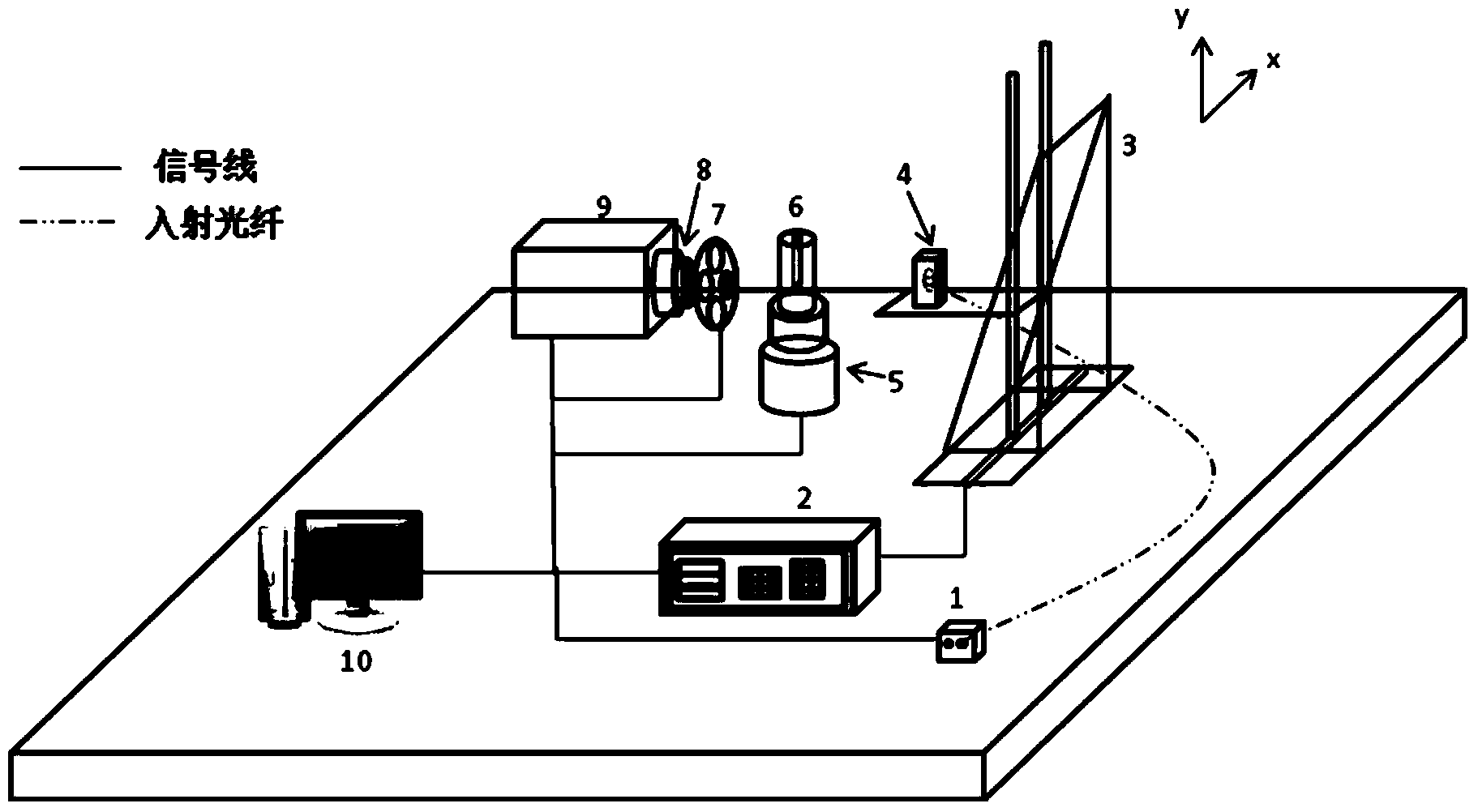
[0037] The system architecture targeted by the present invention is as figure 1 As shown, it is mainly composed of light source 1, incident optical fiber 2, electric control box 3, two-dimensional translation stage 4, coupling lens 5, rotating stage 6, cylindrical imaging cavity 7, filter wheel 8, optical lens 9, electron multiplication charge coupled device 10. Composition of computer 11.

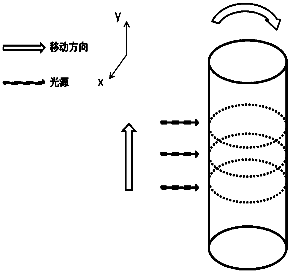
[0038] The invention is aimed at a non-contact small animal FDOT measurement system, which uses a space-scanning laser light source, and the light source emits a steady-state laser with a wavelength of 660nm and a power of 7.5mW. The laser is transmitted to the coupling lens through the incident fiber for beam collimation. The coupling lens installed on the two-dimensional translation stage is controlled by the computer to move in the Y direc...


PUM


Abstract

The invention belongs to the field of medical optical coherent tomography technologies, and relates to a method for extracting information of a fluorescence tomography system for small animals. The method includes calibrating the tomography system and positioning the outer edge of a tomography cavity in a focal plane of a lens; respectively drawing luminous intensity distribution curves of emergent light of excitation light and fluorescence; selecting a range of a central angle theta of the tomography cavity and an information extraction region S mapped to a detected image; individually utilizing pixel data corresponding to a range of small luminous intensity values in the information extraction region S as a sub-region according to the luminous intensity distribution curves of the emergent light, and respectively utilizing pixel data corresponding to two-side ranges of high luminous intensity values as two sub-regions; solving an average value of measured data in each sub-region, utilizing each average value as an average detection value and reconstructing the image by the aid of all the extracted average detection values. The method has the advantages that computation can be simplified, and influence of two-side stray light can be eliminated.

Description

technical field [0001] The invention belongs to the technical field of medical tomography, and relates to an information extraction method for a small animal fluorescence tomography system. Background technique [0002] Fluorescence diffuse optical tomography (FDOT), as a promising small animal imaging[1-2] method, is attracting more and more attention. Its purpose is to monitor the occurrence, metastasis, tumor angiogenesis and antitumor drug treatment response of small animals in real time by irradiating excitation light and measuring the fluorescent signal emitted by the tissue boundary under the calibration of specific fluorescent molecular probes. Non-invasive, specific tracking and detection provide an effective tool for early diagnosis of tumors and detection of subcutaneous tumors in clinical practice in the future. [0003] In recent years, the FDOT system has mostly adopted a non-contact measurement method based on EMCCD (Electron-Multiplying CCD, electron-multipl...

Claims


Application Information

Patent Timeline
Patent Type & Authority Applications(China)
IPC IPC(8): A61B5/00
Inventor 赵会娟王倩周晓青高峰
Owner TIANJIN UNIV
Why Eureka
Try Eureka