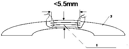
Corneal lamellar piece and production method thereof
A lamellar and corneal technology, used in biology, ophthalmic medicine, and medical fields, can solve the problems of prolapse of eyeball contents, disappearance of cavities, and inability to repair in time, so as to reduce the cost of surgery and solve the problem of excessive waiting time. Effect
- Summary
- Abstract
- Description
- Claims
- Application Information
AI Technical Summary
Problems solved by technology
Method used
Image
Examples
Embodiment 1
[0037] First of all, the patients who underwent full femtosecond laser corneal refractive surgery signed a surgical agreement, and voluntarily donated corneal stroma lenses for clinical treatment. And carry out blood infectious disease inspection, mainly including HIV, HBV, HCV and syphilis and other inspection items. Then, according to the family medical history and social history provided by the patient, unsuitable groups such as drug addicts are excluded.
[0038] Mark the surgical patients who meet the requirements, and collect the corneal stroma lens discarded as medical waste in the full femtosecond laser corneal refractive surgery, mark the corneal stroma lens, and obtain the diameter of the marked corneal stroma lens according to the operation parameter record and the central thickness, the corneal stromal lens that is collected is called the corneal stromal lens unit;
[0039] Record the diameter and central thickness of each corneal stroma lens unit respectively, an...
Embodiment 2
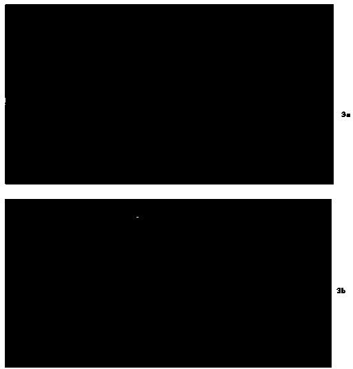
[0054] The repaired patient was a patient with a peripheral corneal perforation. It can be seen that the anterior chamber has disappeared due to the perforation of the cornea in his eye. Its eye perforation is 4.0mm, such as image 3 as shown in a.
[0055] First, the patient's corneal wound was marked with a 4.0mm corneal trephine, and then the necrotic material in the marked ring was removed. Afterwards, the corneal stromal lamina was removed from the glycerin, and the corneal stromal lamella was circumcised with a 4.0mm trephine to form a 4.0mm repair material, which was then sutured to the corneal wound with 10-0 nylon suture.
[0056] After the repair is healed, wait for the visual acuity to improve on its own. After the repair, the OCT image of the eye is as follows: image 3 as shown in b.
[0057]
Embodiment 3
[0059] The repaired patient is a patient with a perforation in the center of the eyeball, and the photo of the eye before repair is as follows Figure 4 as shown in a.
[0060] First, the patient's corneal wound was marked with a 5.5mm corneal trephine, and then the necrotic material in the marked ring was removed. Afterwards, the corneal stromal lamella was removed from the glycerin, and the corneal stromal lamella was cut with a 5.5mm trephine to form a 5.5mm repair material, which was then sutured to the corneal wound with a 10-0 nylon thread to repair the posterior eye photos such as Figure 4 as shown in b.
[0061] After the inflammation disappears, elective optical corneal transplantation can be performed to improve vision.
PUM
 Abstract
Description
Claims
Application Information
 - R&D
- Intellectual Property
- Life Sciences
- Materials
- Tech Scout
- Unparalleled Data Quality
- Higher Quality Content
- 60% Fewer Hallucinations
Browse by: Latest US Patents, China's latest patents, Technical Efficacy Thesaurus, Application Domain, Technology Topic, Popular Technical Reports.
© 2025 PatSnap. All rights reserved.Legal|Privacy policy|Modern Slavery Act Transparency Statement|Sitemap|About US| Contact US: help@patsnap.com


