

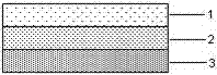
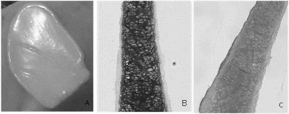
A kind of articular cartilage graft and preparation method thereof
A technology of articular cartilage and grafts, applied in joint implants, joint implants, medical science, etc., can solve the problems of affecting the integration effect, poor biocompatibility, poor long-term effect, etc., and achieve the promotion of extracellular matrix secretion, good pressure resistance and wear resistance, the effect of avoiding immune rejection
- Summary
- Abstract
- Description
- Claims
- Application Information
AI Technical Summary
Problems solved by technology
Method used
Image

Examples
Embodiment 1
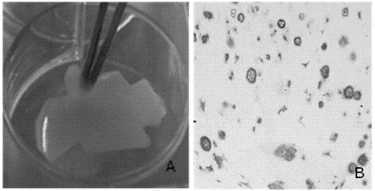
[0030] Step 1: Harvest chondrocytes: cut the superficial cartilage tissue of rabbits into 0.5-3mm 3 Size, first digest with 10 times the volume of 0.1% (w / v) hyaluronidase solution for 30 minutes, shake once every 15 minutes, rinse twice with PBS after digestion; then add 10 times the volume of 0.2% (w / v ) type II collagenase solution for 4 hours, stop the digestion with PBS and fully blow the cells, filter through a 200-mesh sieve, wash with PBS twice, and separate the primary superficial chondrocytes, according to the cell density 1×10 5 piece / cm 2 Inoculate in a culture bottle, add culture solution A, 37°C, 5% CO 2 After culturing in the incubator for 3 days, subculture superficial chondrocytes when they reached more than 80% confluence, add 10 times the volume of 0.25% (w / v) trypsin solution to digest for 3 minutes, stop with PBS and wash 2 times, according to the cell density 1×10 4 piece / cm 2 Inoculate, add culture medium A, and subculture once every 3 days;
[0031]...
Embodiment 2
[0043] Step 1. Acquisition of chondrocytes: cutting superficial porcine cartilage tissue into 0.5-3mm 3 Size, first digest with 10 times the volume of 0.15% (w / v) hyaluronidase solution for 45 minutes, shake once every 15 minutes, rinse twice with PBS after digestion; then add 10 times the volume of 0.25% (w / v) v) Type Ⅱ collagenase solution was digested for 8 hours, PBS was terminated and fully pipetted, filtered through a 200-mesh sieve, washed twice with PBS, and primary superficial chondrocytes were separated, and the cell density was 5×10 5 piece / cm 2 Inoculate in a culture bottle, add culture solution A, 37°C, 5% CO 2 After 7 days of culture in the incubator, when the chondrocytes reached more than 90% confluence, they were passaged, digested with 10 times the volume of 0.5% (w / v) trypsin solution for 8 minutes, terminated with PBS and washed twice, according to the cell density 1×10 5 piece / cm 2 Inoculate, add culture medium A, subculture once every 7 days, and chang...
PUM
| Property | Measurement | Unit |
|---|---|---|
| thickness | aaaaa | aaaaa |
| thickness | aaaaa | aaaaa |
| thickness | aaaaa | aaaaa |
Abstract
Description
Claims
Application Information