Composite 3D printing porous metal support for demineralized bone matrix and preparation method of metal support
A decalcified bone matrix and 3D printing technology, applied in the field of biomedical materials, can solve the problems of unfavorable cell adhesion, inability to realize three-dimensional microenvironment cell culture, difficulty in achieving cell growth, matrix secretion and filling, etc., to achieve The effect of maximizing bone ingrowth
- Summary
- Abstract
- Description
- Claims
- Application Information
AI Technical Summary
Problems solved by technology
Method used
Image


Examples
Embodiment 1
[0057] Example 1 Preparation of 3D printed porous metal scaffold of composite decalcified bone matrix
[0058] 1. Preparation of porous titanium alloy scaffold
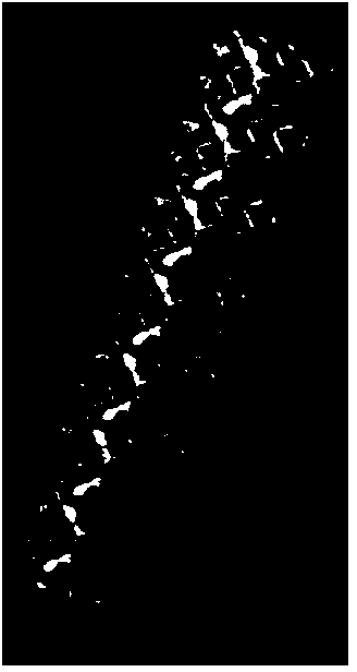
[0059] (1) Import the CT image into three-dimensional image software such as Mimics or CAD to obtain a three-dimensional image of the target bone tissue. The average pore column is 100 μm and the pore diameter is 300 μm. Personalized porous connected 3D digital model (such as figure 1 shown).
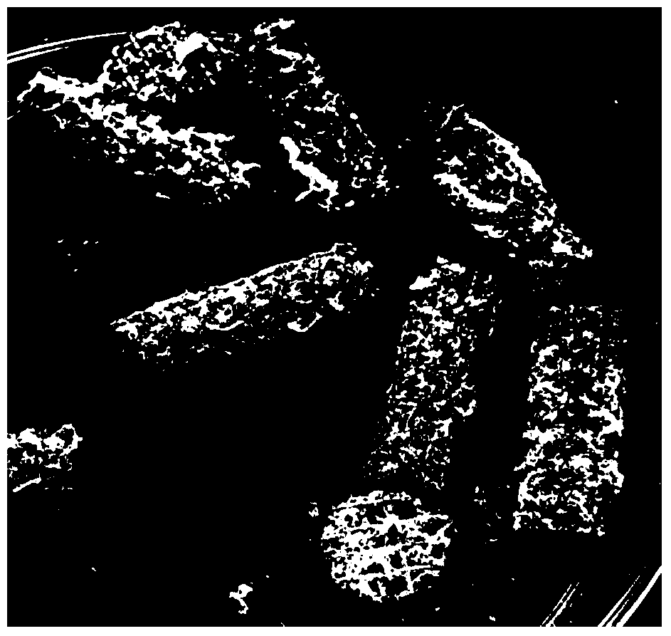
[0060] (2) EOS M280 metal material 3D printer was used to print porous titanium scaffolds (such as figure 2 shown).
[0061] 2. Preparation of decalcified bone matrix
[0062] (1) Take some corpses of New Zealand white rabbits, and cut off their limbs;
[0063] (2) Separate the bones and tissues of the limbs, and remove the periosteum, cartilage and tissues on the surface;
[0064] (3) The obtained limb bones were washed and stored in a -80°C refrigerator for later use;
[0065] (4) Take some New Zealand white rabbit li...
Embodiment 2
[0077] Example 2 Preparation of 3D printed porous metal scaffold of composite decalcified bone matrix
[0078] 1. Preparation of porous titanium alloy scaffold
[0079] (1) Import the CT image into three-dimensional image software such as Mimics or CAD to obtain a three-dimensional image of the target bone tissue. The average pore column is 300 μm and the pore diameter is 1000 μm. Personalized porous connected 3D digital model (such as figure 1 shown).
[0080] (2) EOS M280 metal material 3D printer was used to print porous titanium scaffolds (such as figure 2 shown).
[0081] 2. Preparation of decalcified bone matrix
[0082] (1) Take some corpses of New Zealand white rabbits, and cut off their limbs;
[0083] (2) Separate the bones and tissues of the limbs, and remove the periosteum, cartilage and tissues on the surface;
[0084] (3) The obtained limb bones were washed and stored in a -80°C refrigerator for later use;
[0085] (4) Take some New Zealand white rabbit l...
Embodiment 3
[0097] Example 3 Preparation of 3D printed porous metal scaffold of composite decalcified bone matrix
[0098] 1. Preparation of porous titanium alloy scaffold
[0099] (1) Import the CT image into three-dimensional image software such as Mimics or CAD to obtain a three-dimensional image of the target bone tissue. The average pore column is 1000 μm and the pore diameter is 3000 μm. Personalized porous connected 3D digital model (such as figure 1 shown).
[0100] (2) EOS M280 metal material 3D printer was used to print porous titanium scaffolds (such as figure 2 shown).
[0101] 2. Preparation of decalcified bone matrix
[0102] (1) Take some corpses of New Zealand white rabbits, and cut off their limbs;
[0103] (2) Separate the bones and tissues of the limbs, and remove the periosteum, cartilage and tissues on the surface;
[0104] (3) The obtained limb bones were washed and stored in a -80°C refrigerator for later use;
[0105] (4) Take some New Zealand white rabbit ...
PUM
 Abstract
Description
Claims
Application Information
 - R&D
- Intellectual Property
- Life Sciences
- Materials
- Tech Scout
- Unparalleled Data Quality
- Higher Quality Content
- 60% Fewer Hallucinations
Browse by: Latest US Patents, China's latest patents, Technical Efficacy Thesaurus, Application Domain, Technology Topic, Popular Technical Reports.
© 2025 PatSnap. All rights reserved.Legal|Privacy policy|Modern Slavery Act Transparency Statement|Sitemap|About US| Contact US: help@patsnap.com