

Thyroid microsomal antibody detection reagent kit as well as preparation method and application thereof
A technology of thyroid microsomes and kits, which is applied in the direction of biological testing, measuring devices, material inspection products, etc., can solve the problems of rare detection kits, and achieve the effects of long detection time, long storage time, and small system error
- Summary
- Abstract
- Description
- Claims
- Application Information
AI Technical Summary
Problems solved by technology
Method used
Image

Examples
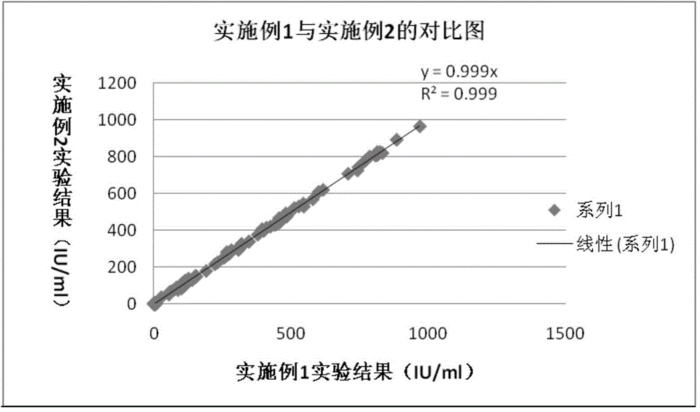
Embodiment 1
[0050] (1) Labeling of protein A
[0051] Preparation of dialysate (F solution): Add Na to a 5000ml beaker 2 CO 3 14.31g, NaHCO 3 26.46g, add purified water to dilute to 4500ml. The prepared F solution was placed on a magnetic stirrer for later use.
[0052] Choose a dialysis bag with a suitable cutoff (usually 14,000), measure the appropriate size, tie one end tightly after wetting, and test for leaks with purified water 3 times (no leakage is required).
[0053] Take 100 μg of protein A and adjust to 1 ml with 0.1 mol / L pH9.5 carbonic acid buffer (F solution). Put it into the dialysate, stir and dialyze at room temperature for 2 hours, add 300 μg of ABEI activated ester to the dialyzed solution, and react at 37°C for 2 hours.
[0054] The ligation product of protein A and ABEI was purified by G-25 gel column.
[0055] D. 2 Solution preparation: add 200ml of 0.5M phosphate buffer (P001 solution), 20g BSA, 8g NaN 3 , 2g MgCl 2 ·6H 2 O, 600ml glycerin, add purified wa...
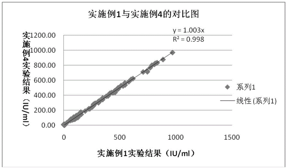
Embodiment 2
[0073] (1) Protein A biotinylation, the specific steps are as follows:
[0074] Preparation of dialysate (F solution): Add Na to a 5000ml beaker 2 CO 3 14.31g, NaHCO 3 26.46g, add purified water to dilute to 4500ml. The prepared F solution was placed on a magnetic stirrer for later use.
[0075] Take 100 μg of biotin and 1 mg of protein A and adjust to 1 ml with 0.1 mol / L pH9.5 carbonic acid buffer (F solution). Put it into the dialysate, stir and dialyze at room temperature for 2 hours, and react at 37°C for 2 hours.
[0076] Purified by G-25 gel column.
[0077] D2 solution was prepared according to the method of Example 1, and the purified connection product was prepared with D 2 The solution was diluted to a concentration of 0.025 µg / ml.
[0078] (2) SA mark
[0079] Take 100μg SA and adjust it to 1ml with 0.1mol / L pH9.5 carbonic acid buffer (F solution). Put it into the dialysate, stir and dialyze at room temperature for 2 hours, add 300 μg of ABEI activated este...
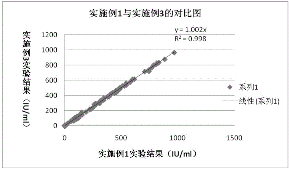
Embodiment 3
[0096] (1) Labeling of protein A
[0097] Preparation of dialysate (F solution): Add Na to a 5000ml beaker 2 CO 3 14.31g, NaHCO 3 26.46g, add purified water to dilute to 4500ml. The prepared F solution was placed on a magnetic stirrer for later use.
[0098] Choose a dialysis bag with a suitable cutoff (usually 14,000), measure the appropriate size, tie one end tightly after wetting, and test for leaks with purified water 3 times (no leakage is required).
[0099] Take 100 μg of protein A and adjust to 1 ml with 0.1 mol / L pH9.5 carbonic acid buffer (F solution). Put it into the dialysate, stir and dialyze at room temperature for 2 hours, add 300 μg of ABEI activated ester to the dialyzed solution, and react at 37°C for 2 hours.
[0100] The ligation product of protein A and ABEI was purified by G-25 gel column.
[0101] D. 2 Solution preparation: add 200ml of 0.5M phosphate buffer (P001 solution), 20g BSA, 8g NaN 3 , 2g MgCl 2 ·6H 2 O, 600ml glycerin, add purified wa...
PUM
| Property | Measurement | Unit |
|---|---|---|
| Particle size | aaaaa | aaaaa |
Abstract
Description
Claims
Application Information