X-ray grating phase-contrast imaging device and method

A phase-contrast imaging and X-ray technology, which is applied in the fields of public security inspection, medical imaging, and non-destructive testing, can solve problems such as the inability to achieve high energy, and achieve large-field phase-contrast imaging, low radiation dose, and high image contrast. degree of effect

Inactive Publication Date: 2015-05-20
UNIV OF SCI & TECH OF CHINA
View PDF4 Cites 23 Cited by
  • Summary
  • Abstract
  • Description
  • Claims
  • Application Information

AI Technical Summary

Problems solved by technology

[0015] The technical problem to be solved by the present invention is that the existing X-ray grating phase contrast imaging device and method cannot realize high energy (>60keV), large field of view (>100×100mm 2 ) imaging, so that it cannot be applied to the bottleneck problem in the fields of clinical medical diagnosis and so on

Method used


Image

  • X-ray grating phase-contrast imaging device and method
  • X-ray grating phase-contrast imaging device and method
  • X-ray grating phase-contrast imaging device and method

Examples


no. 1 example

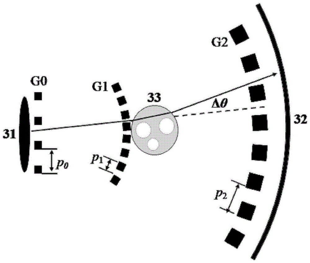
[0060] image 3 It is a schematic diagram of the X-ray grating phase contrast imaging device of the first embodiment and the second embodiment of the present invention. As shown in the figure, the X-ray grating phase contrast imaging device of the present invention includes an X-ray source 31 , a source grating G0 , a beam splitting grating G1 , an analysis grating G2 and a detector 32 . Wherein, the source grating G0 , the beam splitting grating G1 , the analysis grating G2 and the detector 32 are sequentially arranged on the X-ray propagation path of the X-ray source 31 .

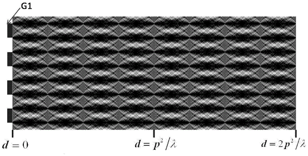
[0061] The imaging principle of the present invention is based on the geometric projection fringe mechanism of the X-ray absorption grating,

[0062] In essence, it is based on the principles of geometric optics, not wave optics. Such as image 3 As shown, the incident X-ray illuminates the beam-splitting grating G1, and in the subsequent geometric projection area, the light intensity distribution appe...

no. 2 example

[0078] The second embodiment of the present invention adopts the same X-ray grating phase contrast imaging device and method as the first embodiment, the difference is that, according to the second embodiment of the present invention, in order to obtain the same information as traditional absorption contrast imaging For the image of the noise ratio, the radiation dose of the X light source 31 of the phase contrast imaging device of the present invention is 20% of the traditional absorption contrast imaging, that is, the radiation dose received by the object is 10% to 20% of the traditional absorption contrast imaging.

[0079] The radiation dose to an object is directly related to the signal-to-noise ratio of the imaging method. One of the advantages of the hard X-ray phase contrast imaging method compared with the traditional absorption contrast imaging method is the improvement of the signal-to-noise ratio of the object image. The signal-to-noise ratio of the object image sa...

no. 3 example

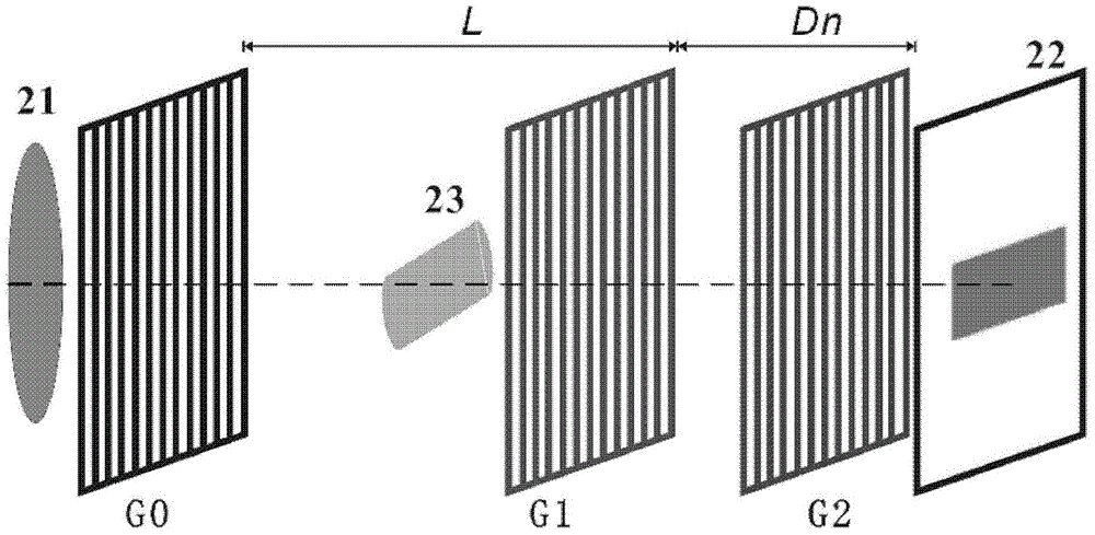
[0088] Figure 4 A third embodiment of the invention is shown. The X-ray grating phase contrast imaging device and method proposed by the present invention are not only applicable to one-dimensional situations, but also can be extended to two-dimensional situations. Such as Figure 4 As shown, on the basis of the first embodiment, both the beam splitting grating G1 and the analysis grating G2 are two-dimensional absorption gratings. The beam-splitting grating G1 and the analysis grating G2 can be obtained by direct fabrication, or by a combination of one-dimensional absorption gratings.

[0089] Similar to the case of the same dimension, the object is placed close to the beam splitting grating G1 to reduce the radiation dose. The refraction and scattering of the X-ray by the object causes the disturbance of the two-dimensional light intensity array in the x and y directions. By analyzing these disturbances, the absorption of the object, the refraction in the x direction, th...


PUM


Abstract

A hard X-ray grating phase-contrast imaging apparatus with large field-of-view, high contrast and low dose and the method thereof. The apparatus includes a source emitter (31), a source grating (G0), a beam splitting grating (G1), an analyzer grating (G2) and a detector (32). The source grating (G0), the beam splitting grating (G1), the analyzer grating (G2) and the detector (32) are orderly arranged in the transmission path of the source emitter (31). The period of the beam splitting grating (G1) is 30-50μm and the aspect ratio is not greater than 20. The apparatus can provide a high image contrast and a low radiation dose and realize a phase-contrast imaging with high energy and a large field-of-view by increasing the grating period, improving the duty ratio of the beam splitting grating (G1) and increasing the distance from an object to the analyzer grating (G2). The apparatus can also utilize the conventional polychromatic x-ray light source and available grating manufacture process, and be suitable for clinical use.

Description

technical field [0001] The present invention relates to technical fields such as medical imaging, non-destructive testing, and public safety inspection, and in particular to an X-ray grating phase contrast imaging device and method, especially a large field of view, high contrast, and low-dose hard X-ray grating Phase contrast imaging device and method. Background technique [0002] In the existing X-ray imaging technology, image contrast comes from the difference of X-ray absorption properties of objects. Therefore, this technology can obtain high image contrast when imaging objects composed of heavy elements such as metals and bones. However, when imaging objects mainly composed of light elements such as carbon, hydrogen, and oxygen (such as human tissue, organic polymer materials, etc.), the image contrast is too low to obtain useful object information. [0003] Compared with traditional absorption contrast imaging, X-ray phase contrast imaging technology can obtain hig...

Claims


Application Information

Patent Timeline
Patent Type & Authority Applications(China)
IPC IPC(8): A61B6/02G01N23/04
CPCA61B6/4035A61B6/4291A61B6/484G01N23/20075G01N23/041
Inventor 吴自玉王志立高昆刘刚潘志云
Owner UNIV OF SCI & TECH OF CHINA
Why Eureka
Try Eureka