

Porcine epidemic diarrhea virus M protein affinity peptides and screening method thereof
A technology for porcine epidemic diarrhea and screening method, which is applied in the field of porcine epidemic diarrhea virus M protein affinity peptide and its screening, can solve the problems of loss of the world pig industry and the failure of vaccination protection, so as to inhibit the replication of the virus, resist the good effect of virus
- Summary
- Abstract
- Description
- Claims
- Application Information
AI Technical Summary
Problems solved by technology
Method used
Image

Examples
Embodiment 1
[0035] Prokaryotic expression of recombinant plasmid pET32a-M:
[0036] According to the published PEDV M gene sequence in GenBank, design primers P1 and P2:
[0037] P1: 5'-CGC GAATTC GCCATGTCTAACGGTTCTAT-3' (the underline indicates that the introduced enzyme cutting site is EcoR I)
[0038] P2: 5'-CGC CTCGAG TTAGACTAAATGAAGCAC-3' (the underline indicates that the introduced restriction site is Xho I)
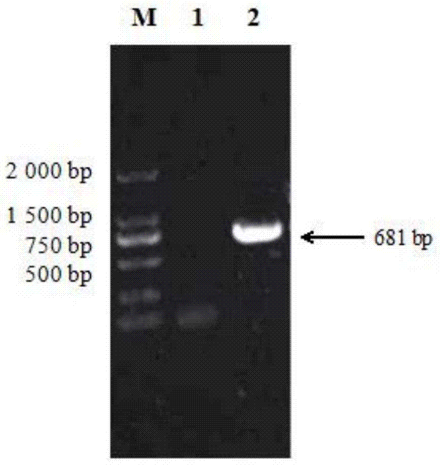
[0039] Select the standard virus strain CV777, extract genomic DNA, and amplify the PEDV M gene fragment by PCR. The amplification conditions are: 94°C pre-denaturation for 10 minutes, 94°C denaturation for 30 seconds, 50.2°C annealing for 30 seconds, 72°C extension for 1 minute, and 94°C pre-denaturation for 10 minutes. Denaturation at 94°C for 30s, annealing at 49.6°C for 30s, extension at 72°C for 1min, a total of 30 cycles, and a final extension at 72°C for 10min. The PCR product was analyzed by agarose gel electrophoresis with a mass fraction of 1%, and there was a ...
Embodiment 2
[0041] Preparation of PEDV M polyclonal antibody:
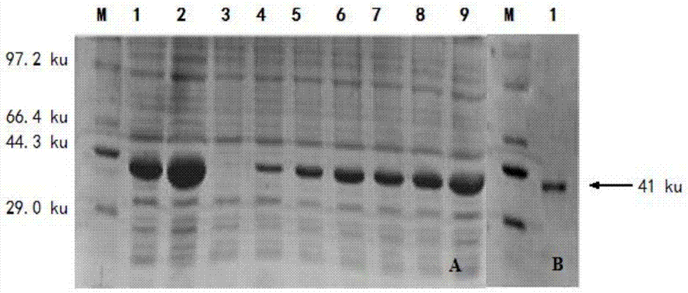
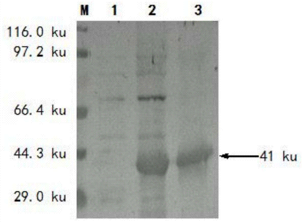
[0042] Select 2-3kg New Zealand white rabbits, the first injection is the purified recombinant M protein and an equal volume of Freund's complete adjuvant after complete emulsification, multi-point subcutaneous injection on the back, the dose is 2 mg / rat, and then the Freund's incomplete adjuvant and equal volume The purified recombinant M protein was completely emulsified and immunized once at intervals of 1 week. After 3 immunizations, the purified recombinant M protein without adjuvant was used to boost the immunization. After 7 days, blood was collected from the rabbit heart to obtain serum, and anti-PEDV M protein polyclonal antibody was obtained. Indirect ELISA was used to detect antibody titers, and Western blot was used to identify the characteristics of polyclonal antibodies. The results of SDS-PAGE analysis showed that a specific band appeared at 41ku after the induction of the recombinant plasmid bacterial liquid, ...
Embodiment 3
[0044] Screening and functional determination of PEDV M affinity peptide:
[0045] (1) Coating of M protein
[0046] The M protein with a concentration of 15 μg / 100 μL was coated on an ELISA plate, blocked overnight at 4°C, and washed 6 times with 0.1% TBST. After washing, add 100 μL of the diluted phage library, shake for 10 minutes, stop for 10 minutes, a total of 40 minutes, wash 10 times with 0.1% TBST, add 100 μL of eluent, shake for 30 minutes, add the supernatant to the EP tube, add 15 μL Neutralizing solution, phage titer determination and amplification, respectively, and subsequent screening. Carry out the next three rounds of screening according to the above steps, and gradually increase the selection pressure, that is, the coating concentration of M protein in each round will be halved sequentially, and the number of TBST washings will be increased by 5 times in each round.
[0047] (2) Amplification of affinity phages: The positive phages after each screening wer...
PUM
 Abstract
Description
Claims
Application Information