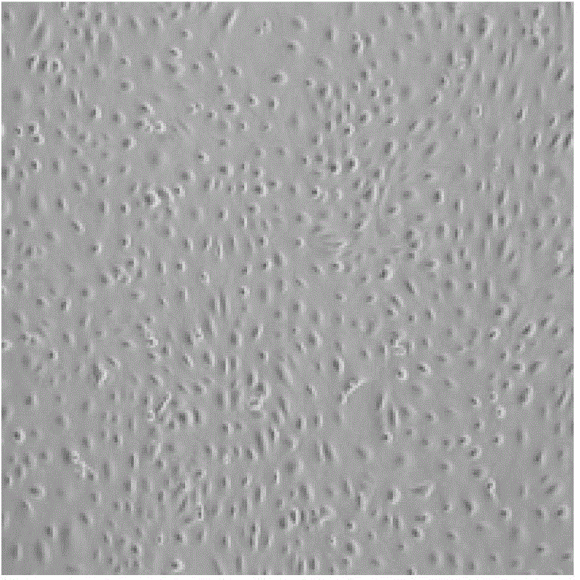
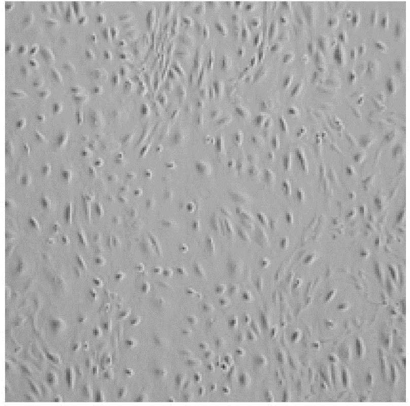
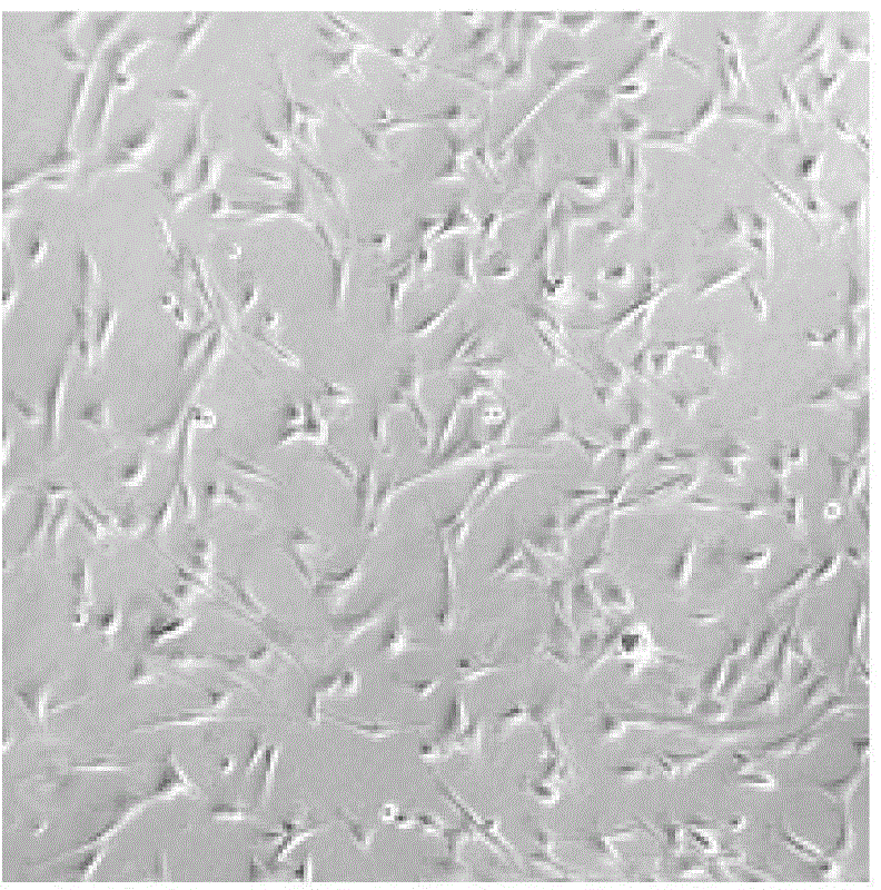
Amnion epithelial cell separation and culture method

A technology of amniotic membrane epithelial cells and amniotic membrane, applied in the field of culture and separation of amniotic membrane epithelial cells, can solve the problems of low purity of amniotic membrane epithelial cells, and achieve the effect of avoiding the reduction of cell adhesion rate, promoting separation and reducing pollution

Active Publication Date: 2015-08-05
天晴干细胞股份有限公司
View PDF5 Cites 7 Cited by
  • Summary
  • Abstract
  • Description
  • Claims
  • Application Information

AI Technical Summary

Problems solved by technology

[0005] The present invention aims to solve the problem of low purity of amnion epithelial cells separated and cultured by existing methods, and provides a method for separating and culturing amnion epithelial cells

Method used


Image

  • Amnion epithelial cell separation and culture method
  • Amnion epithelial cell separation and culture method
  • Amnion epithelial cell separation and culture method

Examples


specific Embodiment approach 1

[0023] Specific embodiment 1: This embodiment is a method for separating and culturing amniotic membrane epithelial cells, which is completed according to the following steps:

[0024] 1. Amniotic membrane tissue separation and disinfection: the amniotic membrane is stripped from the placenta, then washed with saline for 2 to 3 times, and then immersed in saline containing 1% to 3% cefoperazone for 3 to 10 minutes;

[0025] 2. Separation of amniotic membrane epithelial cells: cut the amniotic membrane into 50~150cm 2 Then apply the cut amniotic membrane with the epithelial side up on the sterile nitrocellulose membrane, add 3-10 mL of normal saline to Petri dish A, and then place the amniotic membrane with the epithelial side down on the petri dish In A, let stand for 2~3min to obtain the amniotic membrane patch; place the amniotic membrane patch in petri dish B, add 10-30ml of pancreatin-EDTA with a mass concentration of 0.25% for digestion for 15-20min, and then seal with the para...

specific Embodiment approach 2

[0031] Specific embodiment two: This embodiment is different from specific embodiment one in that the peeling method described in step one is blunt peeling. The other steps and parameters are the same as in the first embodiment.

specific Embodiment approach 3

[0032] Specific embodiment three: This embodiment is different from specific embodiment one or two in that the petri dish A, petri dish B, and petri dish C described in step 2 are petri dishes with a diameter of 9-15 cm. The other steps and parameters are the same as in the first or second embodiment.


PUM

PropertyMeasurementUnit
diameteraaaaaaaaaa

Abstract

The invention relates to an amnion epithelial cell separation and culture method which aims to solve the problem of low purity of the amnion epithelial cells which are separated and cultured by the existing method. The method comprises the following steps: 1. amnion tissue separation and disinfection; 2. amnion epithelial cell separation; and 3. amnion epithelial cell purification culture to obtain the high-purity P0-generation amnion epithelial cell unicell suspension. The amnion patch prepared from the amnion tissues can ensure the flatness of the amnion epithelial surface; meanwhile, the amnion back mucus is closely attached to the connective tissue and nitrocellulose membrane to enhance the trypsinization efficiency and promote the separation between the cells and tissues, so that all the amnion epithelial cells can be collected by short-time digestion, thereby avoiding the reduction of the cell wall-attachment rate; and after the short-time culture, trypsinization is performed twice to obtain the high-purity amnion epithelial cells. The method is applicable to the fields of fundamental research and clinical transplantation.

Description

Technical field [0001] The invention relates to a method for separating and culturing amniotic membrane epithelial cells. Background technique [0002] Amniotic membrane tissue discarded during the perinatal period is one of the important sources of seed cells for regenerative medicine. It has a wide range of sources, is easy to obtain, and has no ethical and moral controversy. It is a research hotspot in recent years. Human amniotic epithelial cells (hAEC) and human amniotic mesenchymal stem cells (hAMSC) can be isolated from the amniotic membrane. Both types of stem cells are rich in content, low in immunogenicity, and non-tumorigenic. The characteristic is the main stem cell resource for clinical treatment in the future, among which amniotic epithelial cells have more "stemness" and are closer to the nature of embryonic stem cells, so they have become a research hotspot in the field of regenerative medicine. [0003] Amniotic membrane epithelial cells develop from the ectoderm ...

Claims


Application Information

Patent Timeline
Patent Type & Authority Applications(China)
IPC IPC(8): C12N5/073
Inventor 付寅生张怡孟庆雪刘艳青
Owner 天晴干细胞股份有限公司
Why Eureka
Try Eureka