Surface-mediated gene therapy type artificial lens and preparation method for same

An intraocular lens and gene therapy technology, applied in intraocular lenses, eye implants, etc., can solve problems such as damage, reduce incidence, avoid toxic side effects, inhibit epithelial cell proliferation or induce lens epithelial cell apoptosis. Effect

Active Publication Date: 2015-08-12
WENZHOU MEDICAL UNIV
View PDF15 Cites 12 Cited by
  • Summary
  • Abstract
  • Description
  • Claims
  • Application Information

AI Technical Summary

Problems solved by technology

[0004] These anti-proliferative drug injections or drug coating methods can better inhibit the proliferation or transdifferentiation of lens epithelial cells, but it is difficult to prevent anti-proliferative drugs from penetrating into the aqueous humor or other ocular tissues, thereby causing damage to other ocular tissues

Method used


Image

  • Surface-mediated gene therapy type artificial lens and preparation method for same
  • Surface-mediated gene therapy type artificial lens and preparation method for same
  • Surface-mediated gene therapy type artificial lens and preparation method for same

Examples


Embodiment 1

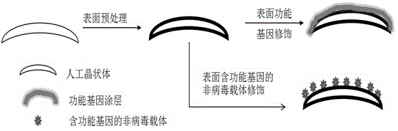
[0027] First, take a clean intraocular lens and surface-treat it with an amino functional group-containing surfactant to bring amine groups on its surface: choose polymethyl methacrylate rigid intraocular lens, immerse into 3mg / mL polyethylene In the imine solution, act for 6 hours, a layer of polyethyleneimine is adsorbed on the surface of the intraocular lens material, washed with water, and dried with nitrogen gas for later use. Then immerse the intraocular lens activated by amine groups on the surface into a 100 μg / mL plasmid DNA solution containing the gene encoding p53 protein, and adsorb a layer of plasmid DNA on the surface of the intraocular lens material through electrostatic adsorption, as shown figure 1 shown.

Embodiment 2

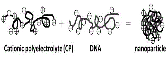
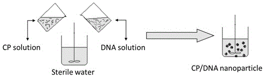
[0029] The cationic polyelectrolyte solution and the plasmid DNA solution containing functional gene fragments are mixed together to prepare electrostatic association nano-gene carriers. The preparation principle is as follows: figure 2 shown. In this embodiment, cationic polyelectrolyte chitosan and plasmid DNA containing a functional gene fragment encoding p53 protein are used as examples. Dissolve 50mg of chitosan (hereinafter abbreviated as CHI) in 50mL of acetic acid buffer solution (pH=5.5). The plasmid DNA (hereinafter abbreviated as pDNA) solution of the fluorescent protein gene was mixed with 10 mL of the above CHI solution, vortexed and oscillated for 30 seconds at room temperature, and the non-viral vector CHI- pDNA nano-associate solution, the preparation process is as follows image 3 shown.

Embodiment 3

[0031]First, take a clean intraocular lens and surface-treat it with an amino functional group-containing surfactant to bring amine groups on its surface: select polymethyl methacrylate rigid intraocular lens, immerse it into 3mg / mL polyethylene In the amine solution, act for 6 hours, a polyethyleneimine layer is adsorbed on the surface of the intraocular lens material, washed with water, and dried with nitrogen gas for later use. Then immerse the intraocular lens activated by amine groups on the surface into 1 mg / mL anionic polyelectrolyte heparin (hereafter abbreviated as HEP) solution, and electrostatically adsorb for 30 minutes, and a layer of negatively charged heparin is adsorbed on the surface of the intraocular lens material; then Immerse the intraocular lens into the non-viral carrier CHI-pDNA nano-association solution containing functional genes prepared in Example 1 for 30 minutes, and a layer of positively charged CHI-pDNA nano-associations is adsorbed on the surfac...


PUM


Abstract

The invention discloses a surface-mediated gene therapy type artificial lens and a preparation method for the same. A plasmid DNA (Deoxyribonucleic Acid) containing a functional gene or a non-viral gene vector loaded with the plasmid DNA containing the functional gene is fixedly arranged on the surface of the artificial lens. The artificial lens and the preparation method for the same have the advantages that a surface-deviated gene therapy vector is obtained by a surface gene modification method, so that the epithelial cell proliferation and transdifferentiation of the lens are suppressed by a surface-mediated gene therapy method, and the artificial lens capable of effectively suppressing after cataract is obtained.

Description

technical field [0001] The invention relates to the field of medical implant materials and device surface modification, in particular to a surface-mediated gene therapy artificial lens and a preparation method thereof. The surface of the intraocular lens of the present invention is immobilized with functional genes, capable of expressing apoptosis protein and / or MicroRNA interfering with cell proliferation, capable of inhibiting lens epithelial cell proliferation or inducing lens epithelial cell apoptosis, and preventing post-cataract occurrence. Background technique [0002] Cataract is the first blinding eye disease in my country. At present, clinical treatment mainly adopts extracapsular phacoemulsification lens extraction combined with intraocular lens implantation. However, various types of intraocular lenses that are widely used at present often face the problem of high incidence of post-cataract after implantation in human eyes. Post-cataract, ie, re-opacification o...

Claims


Application Information

Patent Timeline
Patent Type & Authority Applications(China)
IPC IPC(8): A61F2/16
Inventor 林全愧陈浩
Owner WENZHOU MEDICAL UNIV
Why Eureka
Try Eureka