
Suture-free bilioenteric anastomosis stent manufactured through 3D (three-dimensional) printing and manufacturing method thereof

A 3D printing, biliary and intestinal technology, applied in the field of medical surgical supplies, can solve problems such as affecting tissue blood supply, poor bile duct plasticity, and unsuitability for clinical use, achieving the effect of suture-free anastomosis, saving time, and improving long-term efficacy

Inactive Publication Date: 2015-09-02
洪德飞 +2
View PDF7 Cites 11 Cited by
  • Summary
  • Abstract
  • Description
  • Claims
  • Application Information

AI Technical Summary

Problems solved by technology

Many attempts have been made in this area in previous studies, but so far there is no product that can be used clinically. The reason is that biliary-enteric anastomosis and pancreatico-enteric anastomosis are not like gastrointestinal anastomosis. Because of the strong plasticity of the tissue, the size of the anastomosis The anastomosis is relatively fixed and the anastomosis is large, so staplers have been widely used in clinical practice; the shape, size and caliber of the biliary tract and pancreatic duct vary greatly due to the nature and stage of the disease, and the plasticity of the pancreatic duct and bile duct is poor, and the diameter of the duct is small. Therefore, an anastomotic ring or stapler with the same caliber produced by a fixed mold like a gastrointestinal stapler is not suitable for clinical use.
If it is too thick, it will easily spread excessively and affect the blood supply of the tissue; if it is too thin, it will cause unstable fixation

Method used


Image

  • Suture-free bilioenteric anastomosis stent manufactured through 3D (three-dimensional) printing and manufacturing method thereof
  • Suture-free bilioenteric anastomosis stent manufactured through 3D (three-dimensional) printing and manufacturing method thereof
  • Suture-free bilioenteric anastomosis stent manufactured through 3D (three-dimensional) printing and manufacturing method thereof

Examples


Embodiment Construction

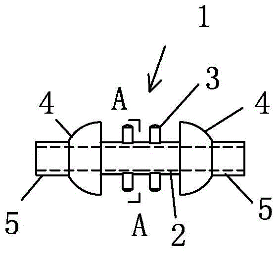
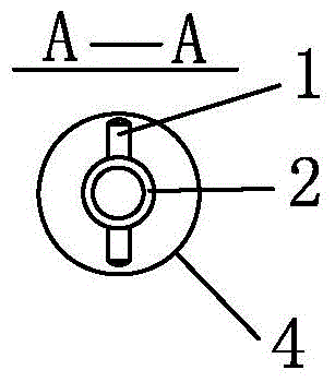
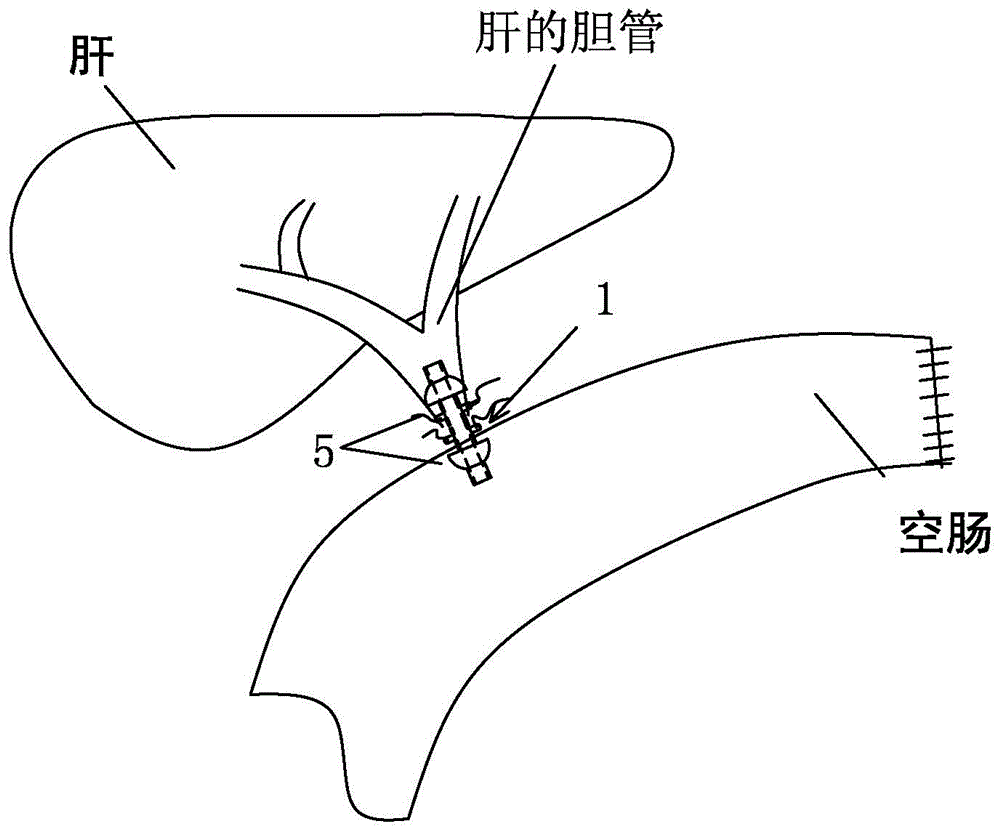
[0021] see figure 1 , figure 2 Shown: a 3D printing suture-free biliary-enteric anastomosis stent, the biliary-intestinal anastomosis stent 1 includes a stent tube 2 and a protruding post 3 protruding from the outer wall of the stent tube, and a convex head is integrally connected to both ends of the stent tube 1 4. A drainage tube 5 is connected to the outer end of the protruding head 4, and the biliary-enteric anastomosis bracket 1 is made of degradable biomaterials by 3D printing technology.

[0022] Wherein, the protruding head 4 is in the shape of a hemisphere, and the arc faces to the outer sides of both ends.

[0023] Wherein, upper and lower protrusions 3 are respectively provided on the outer wall of the bracket tube 2 near the protrusions at both ends.

[0024] A method for preparing a suture-free biliary-enteric anastomosis bracket with 3D printing, comprising the following steps:

[0025] (1) Measure the diameter of the bile duct in patients who need hepatobili...


PUM


Abstract

The invention provides a suture-free bilioenteric anastomosis stent manufactured through 3D (three-dimensional) printing. The bilioenteric anastomosis stent comprises a stent pipe and bulging columns bulging out of the outer wall of the stent pipe, and bulging heads are integrally connected at two ends of the stent pipe. The bilioenteric anastomosis stent is made from a degradable biological material through a 3D printing technique. A manufacturing method comprises steps as follows: (1), the diameter of the bile duct of a patient requiring gallbladder-pancreas reconstructive surgery is tested through CT (computed tomography); (2), required 3D printing data are acquired through algorithm conversion and image processing according to a bile duct CT thin-layer chromatographic scanning result, and the bulging columns and the bulging heads are designed on fit contact surfaces; (3), a 3D printer which is most precise currently is used, the biocompatible degradable material is used, the 3D bilioenteric anastomosis stent is printed and manufactured, and the bilioenteric anastomosis stent is applied to suture-free bilioenteric anastomosis. The suture-free bilioenteric anastomosis stent has the benefits of simple structure, convenience in use, remarkable effect, capability of realizing suture-free bilioenteric anastomosis, the risks of anastomotic leakage and stenosis are reduced, and the operation difficulty and risks are greatly reduced.

Description

technical field [0001] The invention relates to the technical field of medical and surgical supplies, and belongs to a 3D printing suture-free biliary-enteric anastomosis bracket and a preparation method thereof. Background technique [0002] Traditional open pancreatoduodenectomy is complex and traumatic, while biliary-enteric anastomosis and pancreatico-jejunostomy are key steps in pancreaticoduodenectomy. Biliary-enteric anastomotic leakage and pancreaticoenteric anastomotic leakage caused by incomplete healing of mouth) are common serious complications after operation, with a general incidence rate of 5-20%, and the mortality rate of pancreatico-enteric anastomotic leakage can reach 20-40%. Treatment is expensive. [0003] In recent years, a large number of case reports of laparoscopic or da Vinci surgical robotic (minimally invasive) pancreaticoduodenectomy in a few comprehensive large hospitals at home and abroad have shown that minimally invasive pancreaticoduodenect...

Claims


Application Information

Patent Timeline
IPC IPC(8): A61B17/11
CPCA61B17/11A61B17/1114A61B2017/00526A61B2017/00818A61B2017/00831A61B2017/1132
Inventor 洪德飞王知非彭淑牖
Owner 洪德飞
Learn More