A kind of G protein-coupled receptor living body tracing method and application
A technology for coupling receptors and G proteins, applied in chemical instruments and methods, biochemical equipment and methods, preparations for in vivo tests, etc.
- Summary
- Abstract
- Description
- Claims
- Application Information
AI Technical Summary
Problems solved by technology
Method used
Image
Examples
Embodiment 1
[0031] Example 1: Construction of rhodopsin K66M mutant by segmented PCR method
[0032] The purpose of constructing the rhodopsinK66M mutant is twofold, one is to detect whether K66 of rhodopsin is a key residue and whether it can be inserted into eGFP; the other is to introduce a unique Nde I restriction site in rhodopsin to facilitate the insertion of eGFP . The present invention utilizes segmental PCR method to construct rhodopsinK66M mutant, main steps are as follows:
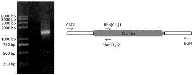
[0033] 1) Using the existing pcDNA4 / TO-rhodopsin plasmid in the laboratory as a template, use the pcDNA4 / TO plasmid general upstream primer CMV and downstream primer Rho(CL1)2 (including the mutation site) to amplify Fragment 1 with a size of about 390bp . The primers used are as follows:
[0034] CMV: 5'-CGCAAATGGGCGGTAGGCGTG-3'
[0035] Rho(CL1)2: 5'CGTGCGCAGCTT CATATG CTGGACGGT-3' (Nde I)
[0036] 2) Using the existing pcDNA4 / TO-rhodopsin plasmid in the laboratory as a template, use the pcDNA4 / TO...
Embodiment 2
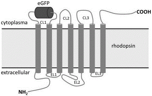
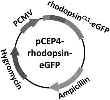
[0055] Example 2: rhodopsin CL1 -eGFP gene construct
[0056] rhodopsin CL1 - The idea of eGFP gene construction is to insert the eGFP gene into pCEP4-rhodopsinK66M to obtain pCEP4-rhodopsin CL1 -eGFP (see figure 2 ).
[0057] Specific steps are as follows:
[0058] 1) Replace the rhodopsinK66M gene fragment from the pCEP4 plasmid to the pBAD24 plasmid: first, pCEP4-rhodopsinK66M and pBAD24 plasmids were digested with BamH I and Hind III, and then the rhodopsinK66M fragment was ligated with the double digested pBAD24 plasmid with T4 ligase Up, the pBAD24-rhodopsinK66M plasmid was obtained.
[0059] 2) Use primers eGFP1 and eGFP2 to amplify the eGFP fragment (about 720bp) with NdeI restriction sites at both ends from the pcDNA3.1-3'-eGFP plasmid, and recover the PCR product from the gel.
[0060] Upstream primer eGFP1: 5'-GGAATTC CATATG GTGAGCAAGGGCGAGGAG-3' (Nde I)
[0061] Downstream primer eGFP2: 5'-CCCTTAAG CATATG CTTGTACAGCTCGTCCAT-3' (Nde I)
[0062] 3) The...
Embodiment 3
[0076] Example 3: pCEP4-rhodopsin CL1 -Expression detection of eGFP in HEK293S cells
[0077] pCEP4-rhodopsin CL1 - The detection of eGFP expression in HEK293S cells mainly includes two aspects, one is to check rhodopsin through Western Blot results CL1 -Expression level and glycosylation processing of eGFP (reference Figure 5 ), the second is to detect rhodopsin by fluorescence microscopy CL1 -Whether eGFP can normally emit green fluorescence and whether it accumulates in cells, etc. (refer to Figure 6 ). The specific operation steps are as follows:
[0078] 1) Plasmid preparation: Prepare the plasmid with a plasmid extraction kit, and select a plasmid concentration of not less than 200ng / μl, A 260 / A 280 High-purity pCEP4-rhodopsin between 1.9 and 2.0 CL1 - eGFP plasmid. The same method was used to prepare pCEP4-rhodopsinWT and pCEP4-rhodopsinK66M plasmids as controls.
[0079] 2) Cell preparation: select HEK293S that has been passaged at least twice after thawin...
PUM
 Abstract
Description
Claims
Application Information
 - R&D
- Intellectual Property
- Life Sciences
- Materials
- Tech Scout
- Unparalleled Data Quality
- Higher Quality Content
- 60% Fewer Hallucinations
Browse by: Latest US Patents, China's latest patents, Technical Efficacy Thesaurus, Application Domain, Technology Topic, Popular Technical Reports.
© 2025 PatSnap. All rights reserved.Legal|Privacy policy|Modern Slavery Act Transparency Statement|Sitemap|About US| Contact US: help@patsnap.com


