Doped self-assembled nano fiber structure and preparation method thereof

A nanofiber and co-assembly technology, applied in the field of biomedical materials and nanomedicine, can solve the problems of clinical application, poor pharmacokinetics and long-term biological safety of photothermal conversion agents, etc., achieve good tumor specificity, and improve tumor accumulation Efficiency and residence time, effect of high tumor accumulation efficiency

Inactive Publication Date: 2015-11-11
XIAMEN UNIV
View PDF1 Cites 5 Cited by
  • Summary
  • Abstract
  • Description
  • Claims
  • Application Information

AI Technical Summary

Problems solved by technology

However, the pharmacokinetics and long-term biological safety of most of the currently reported photothermal conversion agents are poor, so they cannot be used in clinical practice.

Method used


Image

  • Doped self-assembled nano fiber structure and preparation method thereof
  • Doped self-assembled nano fiber structure and preparation method thereof
  • Doped self-assembled nano fiber structure and preparation method thereof

Examples


Embodiment Construction

[0028] Can make the present invention be illustrated more clearly below by specific embodiment:

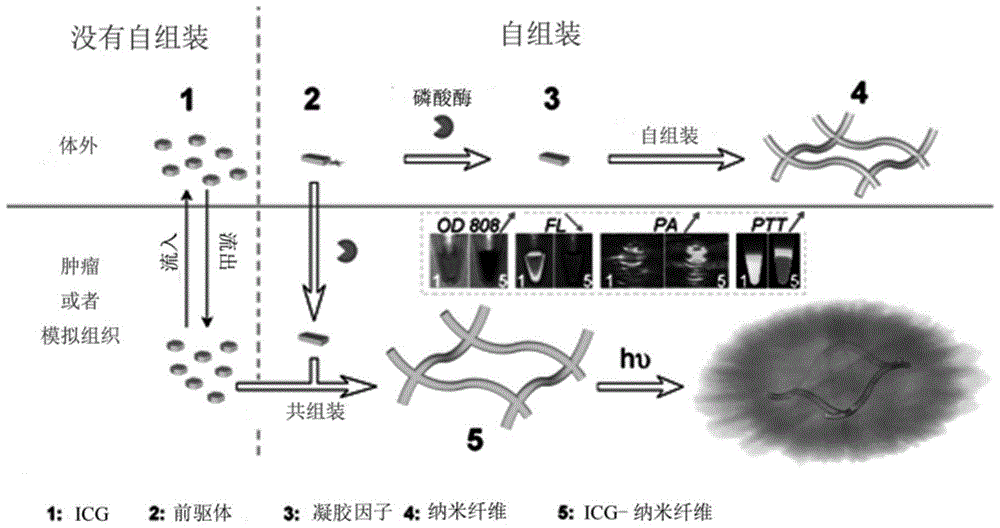
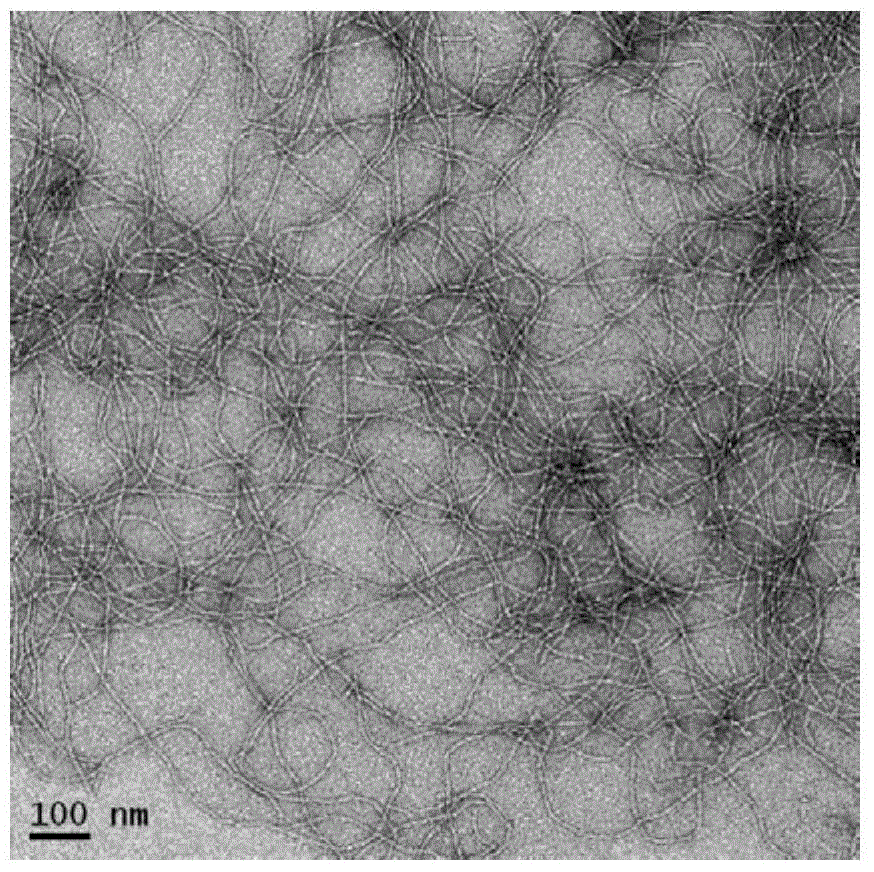
[0029] 1. Preparation of nanofibers:

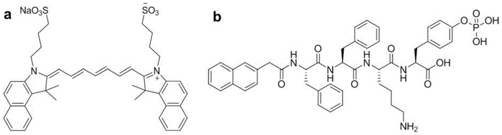
[0030] ICG (10 μg / mL) and NapFFKYp polypeptide (500 μg / mL) were co-dissolved in PBS (PH7-8, 1 mL). ICG-doped nanofibers were formed by adding 1 unit of alkaline phosphatase. Nanofibers without ICG were prepared by the same method as a control.

[0031] 2. Research at the cellular level

[0032] 1) Cultured cells: human-derived cervical cancer cell line HeLa and mouse-derived breast cancer cell line 4T1 were derived from American Type Culture Collection (ATCC), and cultured at 37°C in 5% CO 2 cultured in an incubator.

[0033] 2) Formation of nanofibers in cells: In order to study the formation of nanofibers in cells, 5,000 HeLa cells / well were seeded in a 96-well plate and cultured for 24 hours, and then incubated with different samples: No. 1 sample 10 μg / mL; No. 2 Sample 500 μg / mL; No. 1 sample 10 μg / mL + No. 2 sample 500 μg / mL; No. 1 ...


PUM


Abstract

The invention discloses a doped self-assembled nano fiber structure and a preparation method thereof. Through synergistic interaction of molecules, nano fiber can obviously change near-infrared absorption of a cyanine dye, thereby obviously improving photo-acoustic performance and photo-thermal performance. Phosphatase response synchronous-assembly processes and diagnosis and treatment performance of assembly materials are verified respectively through cell and in vivo experiments. Particularly, through caudal vein injection, after 4 hours, tumors and peripheral normal tissues can be very clearly distinguished. According to the invention, the cyanine dye-doped nano fiber with in situ formation of tumor tissues can be used for fluorescence, photo-acoustic imaging and photo-thermal therapy.

Description

technical field [0001] The invention belongs to the field of biomedical materials and nanomedicine, and specifically relates to a method of using tumor endogenous phosphatase to specifically form an indocyanine green (ICG) doped self-assembled nanofiber structure in tumor tissue, and its preparation method and nanomaterials can be used Integration of diagnosis and treatment in cancer. Background technique [0002] Tumor hyperthermia has become an important tumor treatment method. It uses exogenous stimuli, such as light, radio frequency, microwave, ultrasound and magnetic field, to heat and kill cancer tissue or cancer cells, so as to achieve the purpose of treatment. In particular, photothermal therapy is currently a research hotspot because of its specific selectivity in time and space, and its extremely low invasiveness. During photothermal therapy, the photothermal conversion agent absorbs and converts light energy to generate a lethal temperature in the tumor area. Ho...

Claims


Application Information

Patent Timeline
IPC IPC(8): A61K41/00A61K47/42A61K49/00A61K9/14A61P35/00
Inventor 黄鹏陈小元刘刚楚成超
Owner XIAMEN UNIV
Why Eureka
Try Eureka