Monoclonal antibody for detection and classification of cervical cancer and application thereof
A technology of antibody and detection plate, which is applied in the field of biological diagnosis and medicine, and can solve the problems of culture survival, failure of HPV virus, failure to obtain immune response, etc.
- Summary
- Abstract
- Description
- Claims
- Application Information
AI Technical Summary
Problems solved by technology
Method used
Image
Examples
preparation example Construction
[0181] Preparation of monoclonal antibodies
[0182] Antibodies of the present invention can be prepared by various techniques known to those skilled in the art. For example, an antigen of the invention may be administered to an animal to induce the production of monoclonal antibodies. Monoclonal antibodies can be prepared using hybridoma technology (see Kohler et al., Nature 256; 495, 1975; Kohler et al., Eur.J. Immunol. 6:511, 1976; Kohler et al., Eur.J.Immunol. 6:292, 1976; Hammerling et al., In Monoclonal Antibodies and T Cell Hybridomas, Elsevier, N.Y., 1981), phage display technology or recombinant DNA method (US Patent No. 4,816,567).
[0183]Representative myeloma cells are those that fuse efficiently, support stable high-level production of antibody by selected antibody-producing cells, and are sensitive to culture medium (HAT medium matrix), including myeloma cell lines, such as murine Myeloma cell lines, including those derived from MOPC-21 and MPC-11 mouse tumors...
Embodiment 1
[0215] 1. Preparation of rabbit monoclonal antibody against human papillomavirus HPV16E7
[0216] 1.1 Screening of single-chain antibody (scFv)
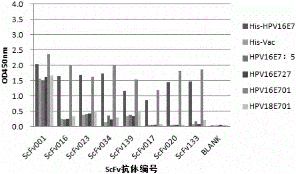
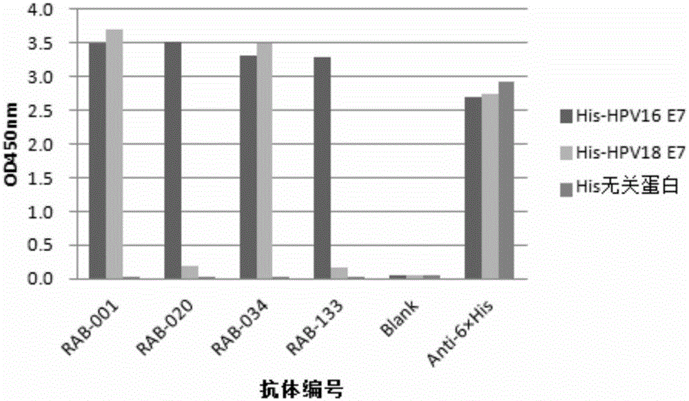
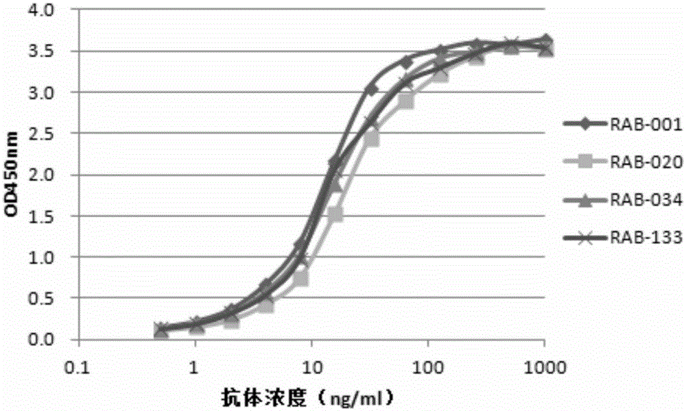
[0217]Rabbits were immunized with His-HPV16E7 recombinant protein, and the titer was detected with His-HPV16E7 recombinant protein and His irrelevant protein. Isolation of rabbit B lymphocytes to obtain immunoglobulin genes. The complete set of variable region genes of B cells is cloned and assembled into a phage antibody library. The constructed phage antibody library was panned with the recombinant protein His-HPV16E7. Enrichment after three rounds of panning; determination of phage titer; amplification of phage plaques; DNA sequencing; Among them, the recombinant protein His-HPV16E7 was selected for ELISA detection and screening, and the negative control (N) was set with His irrelevant protein, the Anti-6×His antibody was set as the positive control (P) coated with His antigen, and the blank control was set at the same time (th...
Embodiment 2
[0235] Example 2 Rabbit-derived monoclonal antibody RAB-034 is used to detect cervical lesion tissue by immunohistochemical staining
[0236] According to the results of antibody screening, rabbit monoclonal antibody RAB-034 was used as the primary antibody to further explore its application in the detection of cervical precancerous lesions. Cervical intraepithelial neoplasia (CIN) is the precancerous lesion of cervical cancer, including cervical mild (CINI grade), moderate (CINII grade), severe dysplasia and carcinoma in situ (CINIII grade). Select normal cervical tissue paraffin sections (negative control), CINI, -II, -III pathological paraffin sections and cervical cancer paraffin sections (positive control), and use RAB-034 to carry out immunohistochemical staining detection, specific operations refer to Example 1, 2.4 section. Results: There were 7 samples of CINI grade, 3 of which had specific staining, and the positive rate was 42.9%; 16 samples of CINII-III grade, 10 ...
PUM
 Abstract
Description
Claims
Application Information
 - R&D
- Intellectual Property
- Life Sciences
- Materials
- Tech Scout
- Unparalleled Data Quality
- Higher Quality Content
- 60% Fewer Hallucinations
Browse by: Latest US Patents, China's latest patents, Technical Efficacy Thesaurus, Application Domain, Technology Topic, Popular Technical Reports.
© 2025 PatSnap. All rights reserved.Legal|Privacy policy|Modern Slavery Act Transparency Statement|Sitemap|About US| Contact US: help@patsnap.com


