Flow Cytometry Assay for Cytotoxic T Cell Degranulation

A flow cytometry and cytotoxic technology, which is applied in individual particle analysis, particle and sedimentation analysis, measurement devices, etc., can solve the problems that the detection results are easily affected by the subjective factors of the examiner, the detection speed is slow, and the accuracy is poor. To achieve the effect of small human factors, high accuracy and fast detection

Active Publication Date: 2018-05-01
倍科为(天津)生物技术有限公司
View PDF3 Cites 0 Cited by
  • Summary
  • Abstract
  • Description
  • Claims
  • Application Information

AI Technical Summary

Problems solved by technology

[0006] The purpose of the present invention is to provide a flow cytometry detection method for cytotoxic T cell degranulation, which can solve the problem of slow detection speed, low precision, poor accuracy and easy detection of detection results in the prior art. The influence of the subjective factors of the tester

Method used


Image

  • Flow Cytometry Assay for Cytotoxic T Cell Degranulation
  • Flow Cytometry Assay for Cytotoxic T Cell Degranulation

Examples


Embodiment 1

[0046] A method for detecting cytotoxic T cell degranulation by flow cytometry, comprising the following steps:

[0047] S11. Separating the peripheral blood mononuclear cells in the sample to be tested and counting to adjust the cell concentration;

[0048] S12, taking the natural target cells of cytotoxic T cells and counting to adjust the cell concentration;

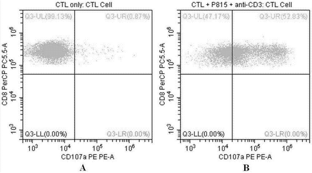
[0049]S13. Mix the peripheral blood mononuclear cells in step S11 and the cytotoxic T cell natural target cells in step S12 in equal proportions by volume and divide them into two groups, and add anti-human CD3 antibody to one of the groups As the experimental group, the other group as the control group; centrifuge the two groups after incubation and discard the supernatant to obtain a precipitate;

[0050] S14. Add flow staining buffer to resuspend the precipitate described in step S13, and add anti-CD3, CD8, D107a flow antibodies and incubate;

[0051] S15. After the incubation is completed, centrifuge, and wash t...

Embodiment 2

[0055] A method for detecting cytotoxic T cell degranulation by flow cytometry, comprising the following steps:

[0056] S21. Separating the peripheral blood mononuclear cells in the sample to be tested and counting to adjust the cell concentration to 1.8×10 6 / ml;

[0057] S22. Take P815 cells, the natural target cells of cytotoxic T cells, count and adjust the cell concentration to 1.8×10 6 / ml;

[0058] S23. Mix the peripheral blood mononuclear cells in step S21 and the cytotoxic T cell natural target cells in step S22 in equal proportions by volume and divide them into two groups, and add anti-human CD3 antibody to one of the groups As the experimental group (the concentration of the antibody is 0.25μg / 100μl), the other group is used as the control group; 2 After incubation in the incubator for 2.5 hours, centrifuge at 1200 rpm for 6 minutes, discard the supernatant, and obtain a precipitate;

[0059] S24. Add flow staining buffer to resuspend the precipitate described...

Embodiment 3

[0065] A method for detecting cytotoxic T cell degranulation by flow cytometry, comprising the following steps:

[0066] S31. Separating the peripheral blood mononuclear cells in the sample to be tested and counting to adjust the cell concentration to 2.2×10 6 / ml;

[0067] S32. Take P815 cells, the natural target cells of cytotoxic T cells, count and adjust the cell concentration to 2.2×10 6 / ml;

[0068] S33. The peripheral blood mononuclear cells in step S31 and the cytotoxic T cell natural target cells in step S32 are mixed in equal proportion by volume and divided into two groups on average, and anti-human CD3 antibody is added to one of the groups As the experimental group (the concentration of the antibody is 0.5μg / 100μl), the other group is used as the control group; 2 After incubation in the incubator for 3.5 hours, centrifuge at 1600 rpm for 4 minutes, discard the supernatant, and obtain a precipitate;

[0069] S34. Add flow staining buffer to resuspend the preci...


PUM


Abstract

The invention relates to the technical field of biology, in particular to a flow-cytometry detecting method of cytotoxic-T-lymphocyte degranulation. The flow-cytometry detecting method includes the steps that cytotoxic-T-lymphocyte degranulation is excited through the antibody-dependent cell-mediated cytotoxic effect, the ratio of the average fluorescence intensity of vesicle-membrane-protein markers CD107a in cytotoxic T lymphocytes after exciting to the average fluorescence intensity before exciting is detected through flow cytometry to evaluate the degranulation function of the cytotoxic T lymphocytes, and if the ratio is larger than or equal to 2.8, the degranulation function is normal. The flow-cytometry detecting method of cytotoxic-T-lymphocyte degranulation is rapid in detection, large in flux, high in accuracy and small in human-factor influence. As key technology details such as natural irritant selecting, the effector target cell ratio and the incubating time are optimized and limited, the stable and normative technical process is built.

Description

technical field [0001] The invention relates to the field of biotechnology, in particular to a method for detecting cytotoxic T cell degranulation by flow cytometry. Background technique [0002] Cytotoxic T-lymphocytes (CTL cells), also known as killer T cells, can specifically kill target cells with antigens, such as transplanted cells, tumor cells, and cells infected by microorganisms, etc., and natural killer cells (Natural killer cells) Killer cell (NK cell) constitutes an important line of defense for the body's anti-virus and anti-tumor immunity. [0003] The most important way for CTL cells to kill target cells-cytolytic killing: After contacting target cells, a series of cytotoxic granule substances (degranulation) can be released, resulting in the lysis of target cells. The detection of degranulation function reflects the killing activity of CTL cells, which is of great significance to the study of cytotoxic function and is an important work in basic medical and c...

Claims


Application Information

Patent Timeline
Patent Type & Authority Patents(China)
IPC IPC(8): G01N15/14
CPCG01N15/14G01N2015/1481
Inventor 王昭
Owner 倍科为(天津)生物技术有限公司
Why Eureka
Try Eureka