Preparation method of small fragment bmg antibody and β2-microglobulin detection kit

A detection kit and microglobulin technology, applied in the field of medical immunology in vitro diagnosis, can solve the problems of expensive imported reagents and low sensitivity, and achieve the effects of increased sensitivity, increased binding rate, and good stability

Active Publication Date: 2019-03-29
上海睿康生物科技有限公司
View PDF11 Cites 0 Cited by
  • Summary
  • Abstract
  • Description
  • Claims
  • Application Information

AI Technical Summary

Problems solved by technology

At present, the β2-microglobulin detection reagents on the market generally have the problem of low sensitivity, and the imported reagents are relatively expensive

Method used


Image

  • Preparation method of small fragment bmg antibody and β2-microglobulin detection kit
  • Preparation method of small fragment bmg antibody and β2-microglobulin detection kit
  • Preparation method of small fragment bmg antibody and β2-microglobulin detection kit

Examples


Embodiment 1

[0033] This embodiment provides a method for preparing a small fragment BMG antibody, the specific steps are as follows:

[0034] Hydrolyze the BMG antibody with protease to obtain Fab fragments;

[0035] Dithiothreitol was added under the condition of pH 8.5, and the disulfide bond of the Fab fragment was opened at room temperature for 5 hours to expose the sulfhydryl group, and the artificial polypeptide was used for coupling to obtain the small fragment BMG antibody.

Embodiment 2

[0037] The present embodiment provides a highly sensitive BMG detection kit, composed as follows:

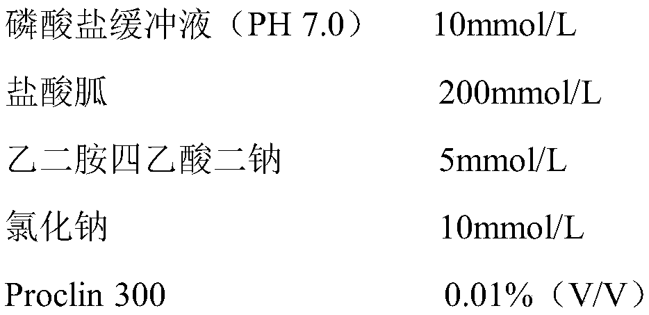
[0038] 1. Reagent R1 is:

[0039]

[0040] 2. The preparation process of reagent R2 is:

[0041] Step 1: Add BMG antibody to latex particles with a particle size of 100nm and incubate at room temperature for 1 hour, wherein the volume ratio of antibody to latex particles is 0.2:1;

[0042] Step 2: Add carbodiimide to the solution obtained in step 1, incubate at room temperature for 2 hours, and add 5 mg of carbodiimide to each milliliter of latex particles;

[0043] Step 3: After centrifuging the suspension solution obtained in Step 2, take the precipitate and wash and disperse it with 10 mmol / L phosphate buffer (pH 7.0).

[0044]The BMG detection kit described in this example is applicable to various types of automatic biochemical analyzers. Taking Hitachi 7170 automatic biochemical analyzer as an example, its operation is shown in Table 1. Analysis method: Two-point endp...

Embodiment 3

[0049] The present embodiment provides a highly sensitive BMG detection kit, composed as follows:

[0050] 1. Reagent R1 is:

[0051]

[0052] 2. The preparation process of reagent R2 is:

[0053] Step 1: Add BMG antibody to latex particles with a particle size of 200nm and incubate at room temperature for 1 hour, wherein the volume ratio of antibody to latex particles is 1:1;

[0054] Step 2: Add carbodiimide to the solution obtained in step 1, incubate at room temperature for 2 hours, and add 5 mg of carbodiimide to each milliliter of latex particles;

[0055] Step 3: After centrifuging the suspension solution obtained in Step 2, take the precipitate and wash and disperse it with 100mmol / L phosphate buffer (PH7..4).

[0056] The specific operation method of the kit is the same as in Example 1.


PUM

PropertyMeasurementUnit
particle diameteraaaaaaaaaa
molecular weightaaaaaaaaaa
concentrationaaaaaaaaaa

Abstract

The invention provides a preparation method of a small-fragment BMG antibody and a high-sensitivity beta2-microglobulin detection kit comprising the antibody. The kit is composed of three parts, namely a reagent R1, a reagent R2 and a calibrator, wherein the reagent R1 is a phosphate buffer system which contains a protein dissociation agent, the reagent R2 is a buffer solution which contains a certain quantity of latex particles coated with the small-fragment BMG antibody, and the calibrator is mainly a bovine serum matrix. By the adoption of the protein dissociation agent, an antigen site in a sample is exposed to the maximum extent, and the combination rate of an antigen and the antibody is increased; the latex particles which are more stable and ideal in signal amplification effect are coated with the special small-fragment BMG antibody; by the two means, high sensitivity of the kit is realized; the problems that BMG reagents on the current market are not high in sensitivity, high in price and the like are solved.

Description

technical field [0001] The invention relates to a preparation method of a small-fragment BMG antibody and a β2-microglobulin detection kit containing the antibody, in particular to the field of medical immune in vitro diagnosis. Background technique [0002] β2-microglobulin was first isolated and discovered by Swedish scholar Berggard in 1968 from the 24-hour urine of patients with renal tubular proteinuria Wilson's disease and chronic cadmium poisoning. It is a serum protein with a molecular weight of 11815Da, and its molecule contains a For disulfide bonds, without sugar, serum protein electrophoresis is located in the β2 region. [0003] The synthesis of β2-microglobulin is relatively stable, and its average production rate is estimated to be 2.4mg / kg / day under normal circumstances. The metabolism of this protein is only dependent on the kidney. There is β2-microglobulin that is shed or released into the blood from the cell surface. It is freely filtered from the glomer...

Claims


Application Information

Patent Timeline
Patent Type & Authority Patents(China)
IPC IPC(8): G01N33/68G01N33/531G01N35/00G01N21/31
CPCG01N21/31G01N33/531G01N33/68G01N35/00
Inventor 李伟奇陈瑛房君江张秀文林清玉
Owner 上海睿康生物科技有限公司
Why Eureka
Try Eureka