PD-1 CAR-T cell as well as preparation method and application thereof

A technology of PD-1CAR-T and PD-1, applied in the field of PD-1CAR-T cells and its preparation, can solve the problems of T cell function exhaustion, T cell death, T cell failure to recognize tumor cells, etc., and achieve high-efficiency tumor The effect of lethal activity

Active Publication Date: 2017-02-15
四川阿思科力生物科技有限公司
View PDF3 Cites 22 Cited by
  • Summary
  • Abstract
  • Description
  • Claims
  • Application Information

AI Technical Summary

Problems solved by technology

However, tumors mainly achieve immune escape from two aspects: (1) The mechanism of tumor cell antigen presentation will be down-regulated or even lose this ability (HLA negative), resulting in the inability of T cells to recognize tumor cells
(2) Many tumor cells express abnormally high PD-L1 molecules, which activate the PD-1 molecules on the surface of T cells, which will lead to the exhaustion of T cell functions and even the death of T cells

Method used


Image

  • PD-1 CAR-T cell as well as preparation method and application thereof
  • PD-1 CAR-T cell as well as preparation method and application thereof
  • PD-1 CAR-T cell as well as preparation method and application thereof

Examples


Embodiment 1

[0029] Lentiviral expression vector preparation

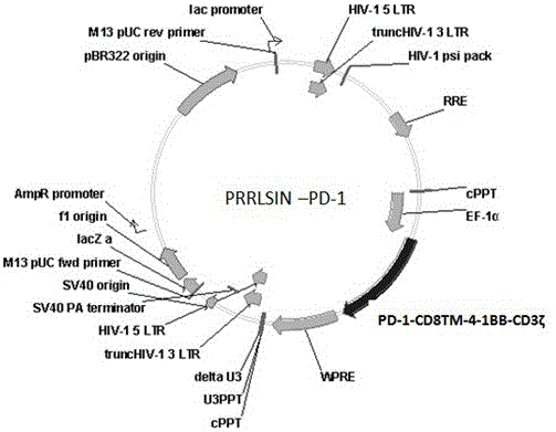
[0030] Gene synthesis PD-1-CD8 TM - The 4-1BB-CD3ζ fusion gene sequence is connected to the PRRSLIN vector through restriction transformation, and the upstream of the gene is the EP-1α promoter. Transform Stbl3 Escherichia coli strain with the vector, screen with ampicillin, obtain positive clones, extract plasmids, identify clones by enzyme digestion, and obtain PRRSLIN-PD-1 lentiviral transfection vector. See the vector construction diagram figure 1 .

Embodiment 2

[0032] lentiviral preparation

[0033] (1) 24 hours before transfection, use about 8×10 per dish 6 293T cells were seeded into 15cm culture dishes. Make sure that the cells are at about 80% confluence and evenly distributed in the culture dish during transfection.

[0034] (2) Prepare solution A and solution B

[0035] Solution A: 6.25 ml 2 × HEPES buffer (the amount packed in 5 large dishes, the effect is the best).

[0036] Solution B: Aliquot and add the following plasmid mixture: 112.5 ug pRRLSIN-EF-PD1 (targetplasmid); 39.5 ug pMD2.G (VSV-G envelope); 73 ug pCMVR8.74 (gag, pol, tat, rev); 625 μl 2M calcium ion solution. Total volume of solution A: 6.25ml.

[0037] Mix solution B well, and while vortexing solution A gently, add solution A drop by drop and let it stand for 5-15 minutes. Gently vortex the above mixed solution of A and B, add dropwise to the culture dish containing 293T cells, gently shake the culture dish back and forth to make the mixture of DNA and c...

Embodiment 3

[0039] Preparation of PD-1 CAR-T cells

[0040] Take 0.5ml of blood for rapid detection of pathogenic microorganisms to exclude microbial infections such as HBV, HCV, HDV, HEV, HIV-1 / 2, Treponema pallidum and parasites; under sterile conditions, use a heparin bottle to collect 50ml of blood (heparin anticoagulant) , immediately (4°C, within 24 hours) to the cell preparation laboratory to ensure that there is no pathogenic microorganism contamination in this process. After obtaining the patient's blood, in the GMP preparation room, wipe the surface of the heparin bottle with an alcohol cotton ball for disinfection and put it into a biological safety cabinet. Open two 50ml centrifuge tubes in advance, transfer the blood into two 50ml centrifuge tubes, and screw them tightly. Put the two 50ml centrifuge tubes filled with blood into the centrifuge for centrifugation. Centrifuge at 400 g (2000 rpm) for 10 min at room temperature to collect the upper layer of plasma, leaving the p...


PUM


Abstract

The invention discloses a PD-1 CAR-T cell as well as a preparation method and an application thereof. Through a transformation method of modifying the T cell by virtue of a chimeric antigen receptor, PD1CD8TM41BBCD3[zeta] molecules are expressed in the T cell. The CAR-T cell prepared by the method can specifically recognize and bind tumor cells which achieve high-expression in PDL-1 protein; therefore, the CAR-T cell can be applied to the preparation of medicines for preventing and treating tumor diseases.

Description

technical field [0001] The present invention relates to the field of cell medicine for tumor treatment, in particular to a PD-1 CAR-T cell and its preparation method and application. Background technique [0002] With the gradual progress of tumor immunotherapy research, programmed death growth factor-1 (PD-1 / CD 279 ) and its ligand PD-L1 / 2 (B7-H1 / CD274) have been acquired as important members of the tumor microenvironment. favored by many researchers. On September 4, 2014, the U.S. Food and Drug Administration (food and drug adm inistration, FDA ) approved Keytruda (pembrolizum ab ) for the treatment of patients with advanced or unresectable melanoma who have not responded to other drug treatments, becoming an obstacle It is the first FDA-approved drug to block the PD-1 cell pathway. PD-1 was first discovered in 1992, and it is mainly expressed in T cells, regulatory T cells, "exhausted" T cells, B cells, and activated cells. Nuclear cells, dendritic cells, natural killer...

Claims


Application Information

Patent Timeline
Patent Type & Authority Applications(China)
IPC IPC(8): C12N5/10C12N15/62C12N15/867A61K35/17A61P35/00
CPCA61K35/17C07K14/7051C07K14/70517C07K14/70521C07K14/70596C12N15/86C07K2319/33C12N2510/00C12N2740/15043C12N15/62C12N2740/16043C07K14/705C07K2319/00A61P35/00C12N5/0636A61K38/00C12N15/85
Inventor 李华顺韩昆昆邓娅任宝永
Owner 四川阿思科力生物科技有限公司
Why Eureka
Try Eureka