
Brain tissue deformation correction system based on wireless transmission

A technology of wireless transmission and brain tissue, which is applied in the field of medical image processing and application, and can solve the problems of not getting rid of dependence, the decrease of accuracy of neurosurgical navigation system, deformation and so on

Inactive Publication Date: 2017-02-22
FUDAN UNIV
View PDF5 Cites 7 Cited by
  • Summary
  • Abstract
  • Description
  • Claims
  • Application Information

AI Technical Summary

Problems solved by technology

However, during the operation, the brain tissue will be deformed due to the influence of gravity, traction or resection, resulting in a decrease in the accuracy of the neurosurgical navigation system based on preoperative images.
[0003] Used in clinical practice, for example, intraoperative magnetic resonance imaging can provide real-time images of intraoperative brain tissue, and is considered an effective method for intraoperative management of brain displacement, but intraoperative magnetic resonance technology is expensive, which limits its use. Clinical application; biomechanical model, using the biomechanical properties of soft tissue to constrain the motion characteristics of soft tissue, and estimating the deformation of the entire brain tissue with the help of finite element equations and intraoperative sparse data, can overcome the shortcomings of intraoperative imaging, Miga[1,2] used the consolidation theoretical model to simulate and correct the deformation of brain tissue after craniotomy, and proved through animal experiments that: the consolidation theoretical model can correct about 75% of the brain tissue deformation error; another 4 clinical cases proved that: after the improvement, the consolidation The average correction rate of the theoretical model can reach 79%. The disadvantage of this method is that it is very difficult to obtain the boundary conditions. This is because the loss of cerebrospinal fluid during the operation is an important parameter for solving the theoretical model of consolidation. That is, to obtain the boundary conditions, it is necessary to It is necessary to know the changes of cerebrospinal fluid after craniotomy, which is also a difficult problem that has been wanted to be solved in clinical practice; Ferrant[3] et al. use linear elastic biomechanical model to model, and use intraoperative magnetic resonance to obtain boundary conditions, adopt this method The surface displacement error of the cerebral cortex is reduced to about 1 mm. The biggest defect of this method is that it does not get rid of the dependence on intraoperative magnetic resonance

Method used


Image

  • Brain tissue deformation correction system based on wireless transmission
  • Brain tissue deformation correction system based on wireless transmission
  • Brain tissue deformation correction system based on wireless transmission

Examples


Embodiment 1

[0051] Embodiment 1 clinical trial

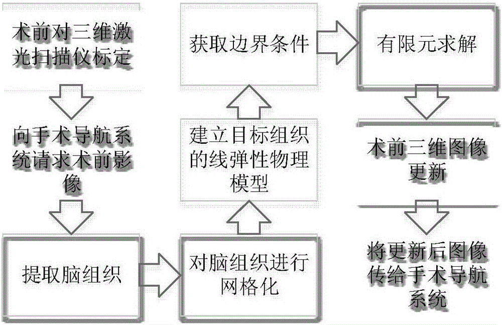
[0052] 1. Install the tracking tool on the 3D laser scanner, and use the calibration module to calibrate to obtain the coordinate transformation relationship from the space transformation of the 3D laser scanner to the space of the tracking tool;

[0053] 2. The brain tissue deformation correction workbench communicates with the neurosurgical navigation system to request the transmission of preoperative image data. The brain tissue deformation correction workbench is the client, and the neurosurgical navigation system is the server. The client first sends the command code CMDASKIMG; the server confirms that the command code is correct, and sends CMDREQACK to confirm; the server sends the preoperative image data to the client, and stores the 240×240×197 three-dimensional MRI brain tissue data in a one-dimensional array; the server Send CMDSENDFIN to stop the transmission; after receiving CMDSENDFIN, the client stops receiving and saves the ...


PUM


Abstract

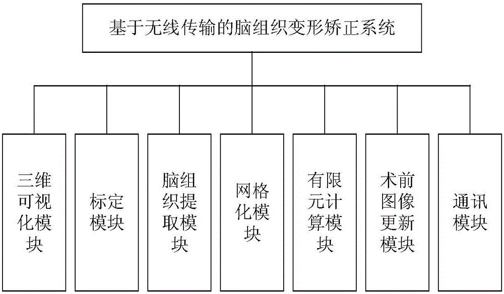
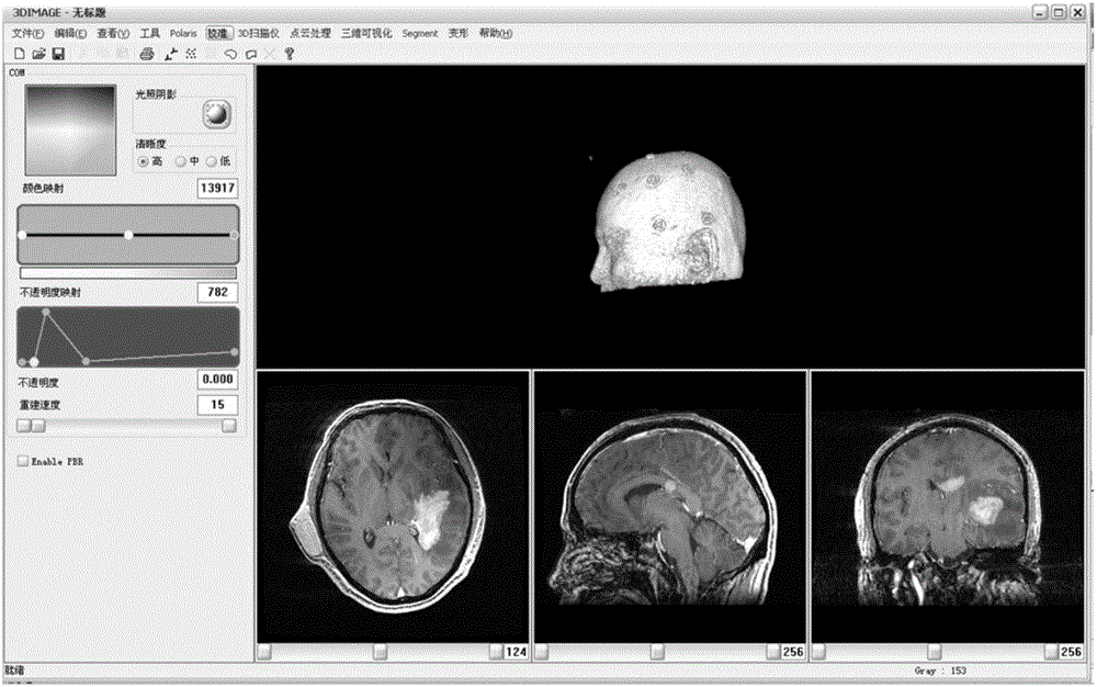
The invention belongs to the fields of medical image processing and application, and relates to a brain tissue deformation correction system based on wireless transmission, in particular to an intraoperative brain tissue deformation correction system applied to neurosurgical department. The system comprises a brain tissue deformation workbench and a three-dimensional laser scanner workbench. A brain tissue deformation correction software system is loaded in the brain tissue deformation correction system workbench, and the brain tissue deformation correction software system can communicate with a neurosurgical operation navigation system by virtue of a wireless local area network. The brain tissue deformation correction software system comprises a three-dimensional visual module, a calibration module, a brain tissue extracting module, a grid module, a boundary condition acquisition module, a finite element calculating module, a preoperative image updating module and a communication module. The system is reliable in precision; the system can be integrated in the existing neurosurgical operation navigation system, and the system is conducive to the implementation of intraoperative soft tissue deformation correction, so that the precision of the navigation system is greatly improved, and the system is beneficial for clinical application.

Description

technical field [0001] The invention belongs to the field of medical image processing and application, and relates to a brain tissue deformation correction system based on wireless transmission, in particular to a brain tissue deformation correction system in neurosurgery. The system includes a brain tissue deformation workbench and a three-dimensional laser scanner workbench, which can be integrated into the existing neurosurgery navigation system to help achieve intraoperative soft tissue deformation correction, thereby greatly improving the accuracy of the navigation system and contributing to clinical applications. Background technique [0002] The prior art discloses that a neurosurgery navigation system (Image guided Neurosurgery System, IGNS) is helpful to improve the quality of surgical operation and reduce brain injury during operation. However, during the operation, the brain tissue will be deformed due to the influence of gravity, traction or resection, resulting ...

Claims


Application Information

Patent Timeline
IPC IPC(8): A61B34/20A61B34/10
Inventor 章琛曦宋志坚姚德民史勇红王满宁葛炜高立希
Owner FUDAN UNIV
Learn More