
Radiative imaging system with high resolution and imaging method and application thereof

A high-resolution, optical imaging technology, which is applied in the field of biomedical molecular imaging, can solve the problems that radiation imaging cannot image target imaging, limit the clinical transformation of radiation imaging, and cannot accurately image, etc., and achieve good clinical application prospects and scientific research value, biological safety and deep tissue optical signal penetration problems, and the effect of overcoming the problem of poor resolution

Active Publication Date: 2017-03-22
FOURTH MILITARY MEDICAL UNIVERSITY
View PDF5 Cites 1 Cited by
  • Summary
  • Abstract
  • Description
  • Claims
  • Application Information

AI Technical Summary

Problems solved by technology

[0004] However, due to the spatial scattering properties of radioactive radiation, although radiation imaging has the aforementioned advantages and can be used for human body imaging, there is a bottleneck problem of poor resolution. Accurate imaging; at the same time, the deeper the distance between the imaging target and the body surface, the higher the degree of ray scattering and the poorer the resolution. Traditional radiation imaging cannot accurately image the deep imaging target
The above two points greatly limit the clinical transformation of radiation imaging

Method used


Image

  • Radiative imaging system with high resolution and imaging method and application thereof
  • Radiative imaging system with high resolution and imaging method and application thereof
  • Radiative imaging system with high resolution and imaging method and application thereof

Examples


Embodiment Construction

[0030] The present invention will be described in detail below in combination with specific embodiments.

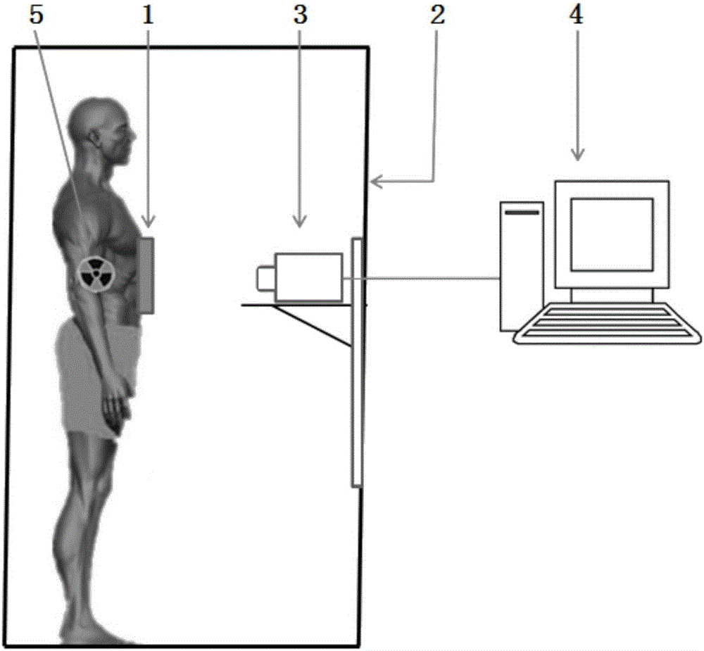
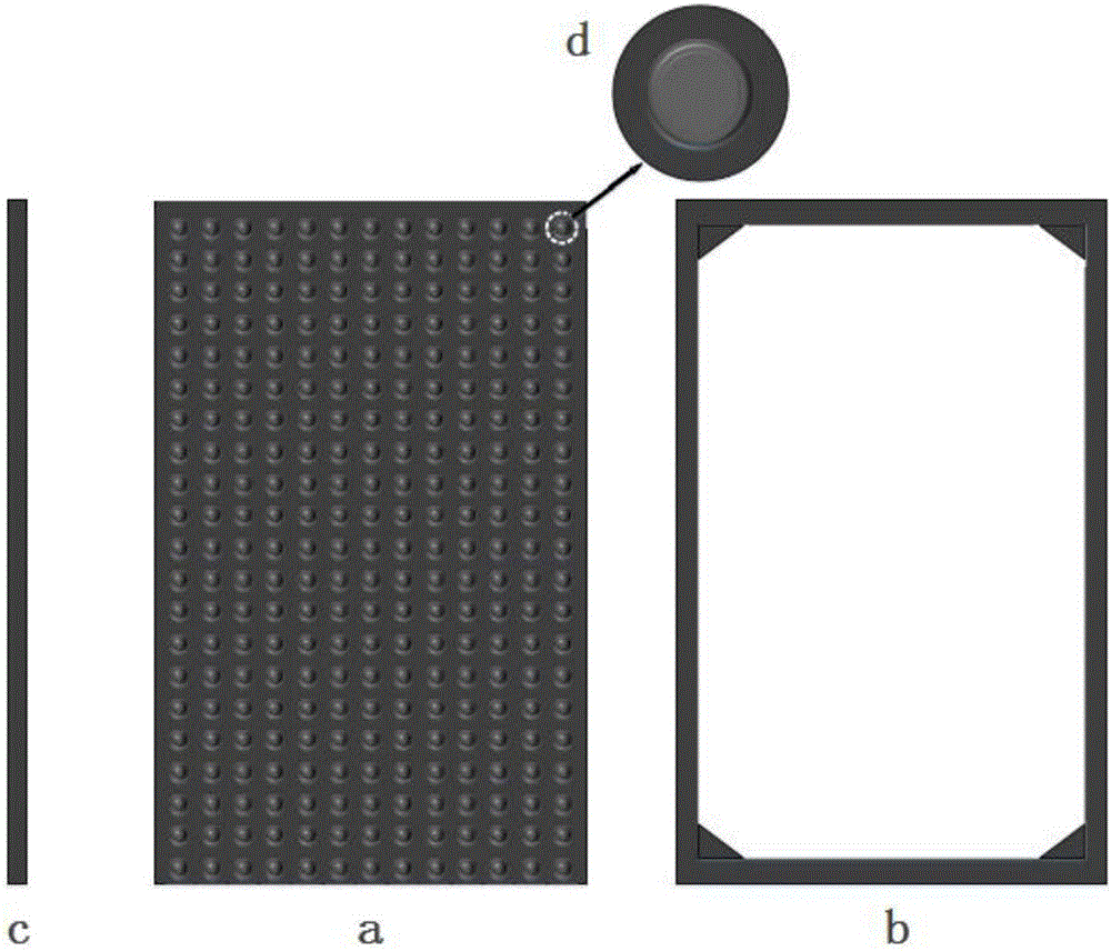
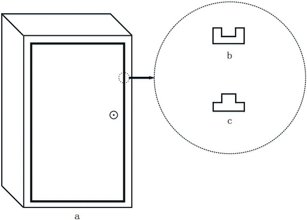
[0031] The present invention is based on the radiation imaging concept and improves and optimizes the radiation imaging imaging system for resolution, which is suitable for accurate imaging of clinical or experimental animals. like figure 1As shown, the radiation imaging system provided by the present invention includes an intensifying screen radiation conversion film 1 (referred to as film), an imaging darkroom 2 and an optical imaging hardware system (comprising an EMCCD camera 3 and a computer 4) with a parallel aperture collimator. ). The film is composed of a clinically used X-ray rare earth intensifying screen (calcium tungstate medium-speed medical intensifying screen) and a parallel-hole collimator made of flexible lead material. The parallel-hole collimator can shield scattered radiation. Increase the display resolution. The imaging darkroom is manufactured ac...


PUM

PropertyMeasurementUnit
Diameteraaaaaaaaaa
Center distanceaaaaaaaaaa
Thicknessaaaaaaaaaa

Abstract

The invention relates to a radiative imaging system. Aiming at the neckbottle problem which exists in the prior art that radiative imaging is not excellent in resolution, the invention provides a radiative imaging system with high resolution and an imaging method and application thereof. The radiative imaging system comprises an intensifying screen radiative converting sticking film for converting a photon emitted by a nuclide into a radiative signal, an imaging darkroom for accommodating a radioactive source and an optical imaging hardware system used for collecting and obtaining a fused image. The imaging method comprises the steps of collecting a white-light image and a radiative image of the radioactive source, conducting similarity and filtration on the radiative image, fusing the radiative image with the white image after adding a pseudo color, and finally providing reliable functional image information. According to the radiative imaging system with high resolution and the imaging method and application thereof, improvement on the resolution of radiative imaging is conducted through a parallel hole collimator in the intensifying screen radiative converting sticking film, the radiative imaging system can be applicable to accurate imaging clinically or for experimental animal, and thus the problems in the prior art of biosecurity and deep tissue optical signal penetrability are effectively solved.

Description

technical field [0001] The invention relates to the technical field of biomedical molecular imaging, in particular to a high-resolution radiation imaging system and its imaging method and application. Background technique [0002] The clinical applications of optical imaging mainly include Cerenkov imaging (thyroid, axillary lymph nodes) and fluorescence imaging (intraoperative navigation, sentinel lymph nodes). The advantage of Cerenkov light imaging is that it directly uses mature nuclide probes for imaging. It is a natural nuclide-optical multimodal imaging mode with high safety and flexibility. The disadvantage is that the light intensity is weak , poor penetration; fluorescence imaging light intensity is high, but the types of fluorescent imaging agents that can be used clinically are extremely limited, and the excitation light and emission light are still limited by the limited tissue penetration, and non-specific fluorescence will also reduce the signal noise ratio. ...

Claims


Application Information

Patent Timeline
IPC IPC(8): A61B5/00A61B6/00
Inventor 康飞汪静曹旭曹欣马晓伟张惠智
Owner FOURTH MILITARY MEDICAL UNIVERSITY
Learn More