Shortwave infrared fluorescence microimaging method

A short-wave infrared and microscopic imaging technology, used in fluorescence/phosphorescence, material analysis by optical means, measurement devices, etc., to achieve high imaging resolution, broad development and application prospects, and the effect of large biological tissue penetration depth.

Inactive Publication Date: 2017-04-26
ZHEJIANG UNIV
View PDF9 Cites 13 Cited by
  • Summary
  • Abstract
  • Description
  • Claims
  • Application Information

AI Technical Summary

Problems solved by technology

At present, there have been some short-wave infrared fluorescence macroscopic in vivo imaging systems that have been put into use, but there is no method that can provide short-wave infrared fluorescence microscopic imaging.

Method used


Image

  • Shortwave infrared fluorescence microimaging method

Examples


Embodiment Construction

[0012] The present invention will be further described below in conjunction with accompanying drawing.

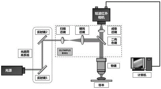
[0013] Such as figure 1 As shown, the short-wave infrared fluorescence microscopy imaging system includes a near-infrared light source (or visible light source), an optical path climbing system, a beam expander system, a beam splitter (dichroic mirror), an objective lens, an imaging lens, a short-wave infrared camera, a computer, etc. .

[0014] First, pass the external near-infrared light source (or visible light source) through an optical path climbing system (composed of two mirrors, such as figure 1 Shown in mirror 1 and mirror 2) introduces the imaging optical path module, which borrows the imaging optical path module in the commercial Olympus BX61 upright microscope imaging system (of course, this module can also be built by itself, or borrowed from other Traditional microscopic imaging system), the excitation light source is expanded by a beam expander system compo...


PUM


Abstract

The invention discloses a shortwave infrared fluorescence microimaging method. A near-infrared light source or a visible light source is used as a light source for exciting a shortwave infrared fluorescence probe, the excitation light source converges on a shortwave infrared fluorescent sample through an objective lens and excites fluorescence, the fluorescence is collected by the objective lens and goes through a beam splitter to separate excitation lights and a fluorescence signal, only the fluorescence signal reaches a shortwave infrared camera, and the shortwave infrared camera transmits a video signal to a computer in order to realize real-time shortwave infrared imaging. Tomographic fluorescence imaging of different depths of the sample can be obtained by adjusting the relative position of the fluorescent sample and the objective lens. Compared with the traditional fluorescence microscopy methods in the visible light band, the method disclosed in the invention has the advantages of large biological tissue penetration depth and small biological damage; and compared with shortwave infrared fluorescence macroimaging methods, the method disclosed in the invention has the advantages of high imaging resolution, high imaging magnification factor, video microimaging realization and tomographic fluorescence imaging realization.

Description

technical field [0001] The invention belongs to the field of microscopic imaging of applied optics, and is a short-wave infrared fluorescence microscopic imaging system (Short-wave Infrared Fluorescence Microscope, SWIRFM for short) which combines traditional fluorescence microscopic imaging technology and short-wave infrared technology. Background technique [0002] Fluorescence microscopy has been widely used in biological research. Compared with traditional transmission microscopy imaging, fluorescence microscopy imaging can provide a higher imaging signal-to-noise ratio, and the use of fluorescent materials for specific labeling of specimens is more conducive to extraction Microscopic information of specific locations. A common fluorescence microscopy imaging system follows the basic reflective fluorescence excitation design, which mainly includes a light source part, a beam splitter part, an objective lens, a fluorescent specimen and an imaging part. The principle is t...

Claims


Application Information

Patent Timeline
Patent Type & Authority Applications(China)
IPC IPC(8): G01N21/64
CPCG01N21/6458
Inventor 钱骏孙超伟张鹤群
Owner ZHEJIANG UNIV
Why Eureka
Try Eureka