
Separation method of mouse intestinal lamina propria primary dendritic cells

A technology of dendritic cells and lamina propria, applied in the field of isolation of primary dendritic cells in the lamina propria of the mouse intestine, can solve problems such as difficulty in achieving satisfactory results, easy death, and excessive cell damage, and shorten the potential Toxic action time, reduce mechanical damage, and increase cell productivity

Active Publication Date: 2017-05-31
NANJING DRUM TOWER HOSPITAL
View PDF3 Cites 2 Cited by
  • Summary
  • Abstract
  • Description
  • Claims
  • Application Information

AI Technical Summary

Problems solved by technology

The disadvantages of this method include: ① excessive mechanical damage caused by repeated shearing of the small intestine; ② cells in the epithelial layer, lamina propria, and serosa layer were not digested one by one, and mixed digestion would inevitably cause the cells in the epithelial layer and serosa layer to interfere with each other. The interference of lamina propria cells is too large, and the purity of lamina propria cells is significantly reduced; ③dendritic cells are sensitive, fragile, and easy to die. Therefore, it is difficult to achieve satisfactory results

Method used


Image

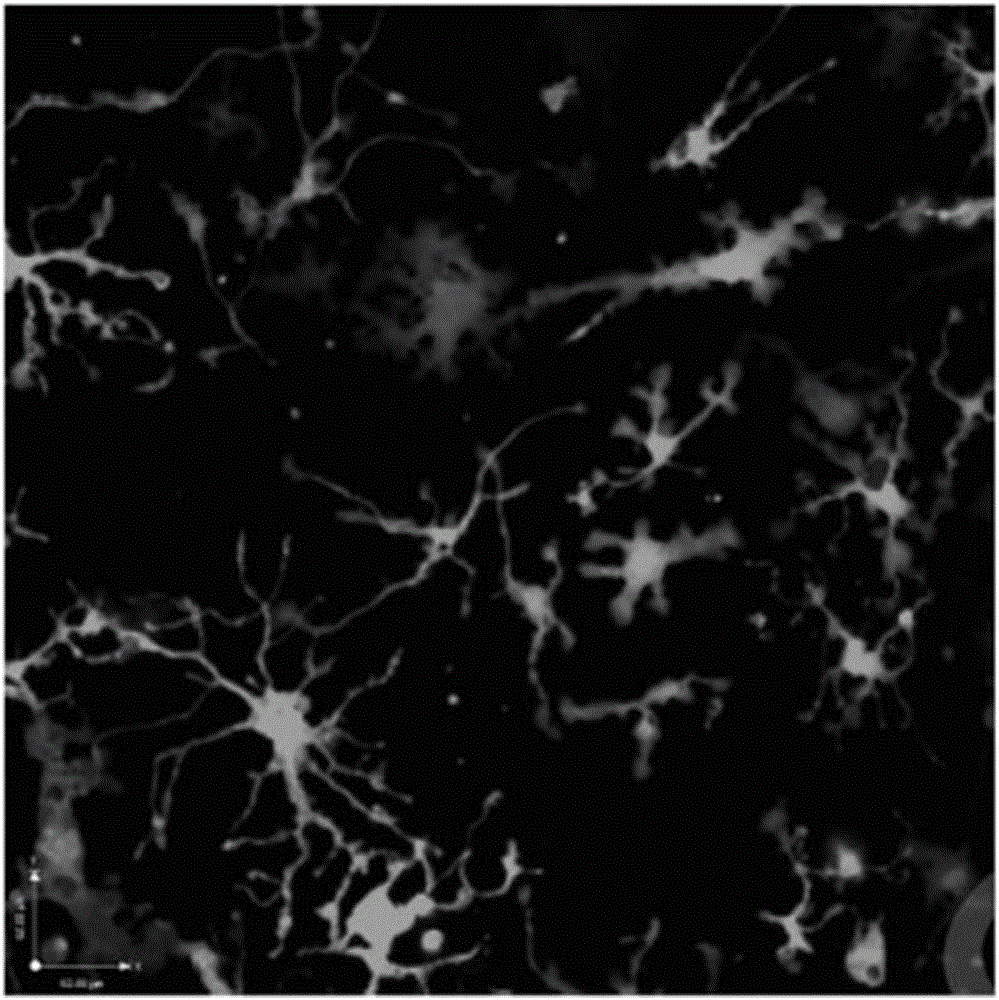
  • Separation method of mouse intestinal lamina propria primary dendritic cells
  • Separation method of mouse intestinal lamina propria primary dendritic cells
  • Separation method of mouse intestinal lamina propria primary dendritic cells

Examples


Embodiment 1

[0032] 1. Put mice in routine CO 2 After execution, the abdominal wall and peritoneum were cut longitudinally with scissors to expose the abdominal viscera. The scissors intercepted the entire small intestine from the duodenum to the ileocecal, and put it in balanced salt solution A for temporary storage;

[0033] 2. Cut the small intestine into 4 segments on average (each segment is about 10-12cm long), ligate one end of the intestinal segment with silk thread, turn the intestinal segment to the surface of the thin tube with a polyethylene thin tube (so that the inner surface of the intestinal tube is exposed), and fix it with silk thread ligation (Such as figure 1 ), put into the 15ml test tube that 12ml balanced salt solution B is housed;

[0034] 3. After manually shaking the test tube for 2 minutes, replace it with a new 12ml balanced salt solution B, and shake it for another 2 minutes. Repeat this for a total of 3 times to wash away impurities such as fecal residue on t...


PUM


Abstract

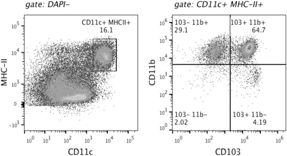
The invention discloses a separation method of mouse intestinal lamina propria primary dendritic cells. The method comprises the following steps of cutting intestinal canals into intestinal sections being 10 to 12cm; eluting intestinal canal surface impurities and intestinal canal surface mucus; using digestive juice B containing EDTA (ethylene diamine tetraacetic acid) and EGTA (ethylene glycol tetraacetic acid) for eluting the enterocyte layer; then, performing digestion by digestive juice containing Liberase TL and DNase I; finally, performing sorting by a flow cytometry to obtain the mouse intestinal lamina propria primary dendritic cells. The primary dendritic cells obtained through the separation by the method have high purity and complete phenotype.

Description

technical field [0001] The invention belongs to the field of biology, and relates to a method for isolating primary dendritic cells of the intestinal lamina propria of mice. Background technique [0002] The intestinal tract is mainly composed of epithelial layer, lamina propria and serosal layer. The epithelial layer is mainly a layer of flat epithelial cells, while the lamina propria contains a large number of immune cells, which are recognized and captured by mammals entering through the digestive tract. An important place for external pathogenic microorganisms in the body, the immune disorder of the intestinal lamina propria is closely related to various diseases such as infection, tumor, and inflammatory bowel disease. Dendritic cells are one of the most important immune cells. Research on dendritic cells will help improve the clinical diagnosis and treatment level of anti-infection and anti-tumor, and improve the prognosis of patients. The isolation and purification o...

Claims


Application Information

Patent Timeline
IPC IPC(8): C12N5/0784
CPCC12N5/0639C12N2509/00
Inventor 刘颂管文贤任建安王革非王萌汪灏
Owner NANJING DRUM TOWER HOSPITAL
Learn More