X-ray, ultrasonic and infrared image fusion-based breast mass image detection method

An image fusion and image detection technology, applied in the field of image processing, can solve problems such as single source of breast image information, inability to conveniently view breast images, and insufficient image information richness, to achieve accurate diagnostic information, good denoising effect, and reliability and the effect of increased effectiveness

Inactive Publication Date: 2017-05-31
XIDIAN UNIV
View PDF4 Cites 15 Cited by
  • Summary
  • Abstract
  • Description
  • Claims
  • Application Information

AI Technical Summary

Problems solved by technology

However, the existing methods are still based on a pair of breast images. The information source of breast images is single, and the richness of image information is not enough to obtain more accurate information.
Moreov

Method used


Image

  • X-ray, ultrasonic and infrared image fusion-based breast mass image detection method
  • X-ray, ultrasonic and infrared image fusion-based breast mass image detection method
  • X-ray, ultrasonic and infrared image fusion-based breast mass image detection method

Examples


Embodiment 1

[0027] In recent years, with the development of science and technology, the diagnostic techniques and treatment methods of breast cancer have been greatly improved. In terms of diagnostic techniques, the more common methods mainly include mammography soft x-ray examination, ultrasound imaging examination, thermal image examination, near-infrared scanning examination, CT examination, tumor marker examination and biopsy and other methods; in terms of treatment methods , has changed from the previous surgical treatment-based method to comprehensive treatment-based methods, such as endocrine therapy, CSRT treatment, cell therapy, gene therapy, etc. Although people have made outstanding progress in the treatment of breast cancer, they can only achieve good results in the early treatment of cancer, because most advanced cancers have metastasized to distant places, no matter what modern treatment measures are used, Curative effect is still unsatisfactory. Early detection and diagnos...

Embodiment 2

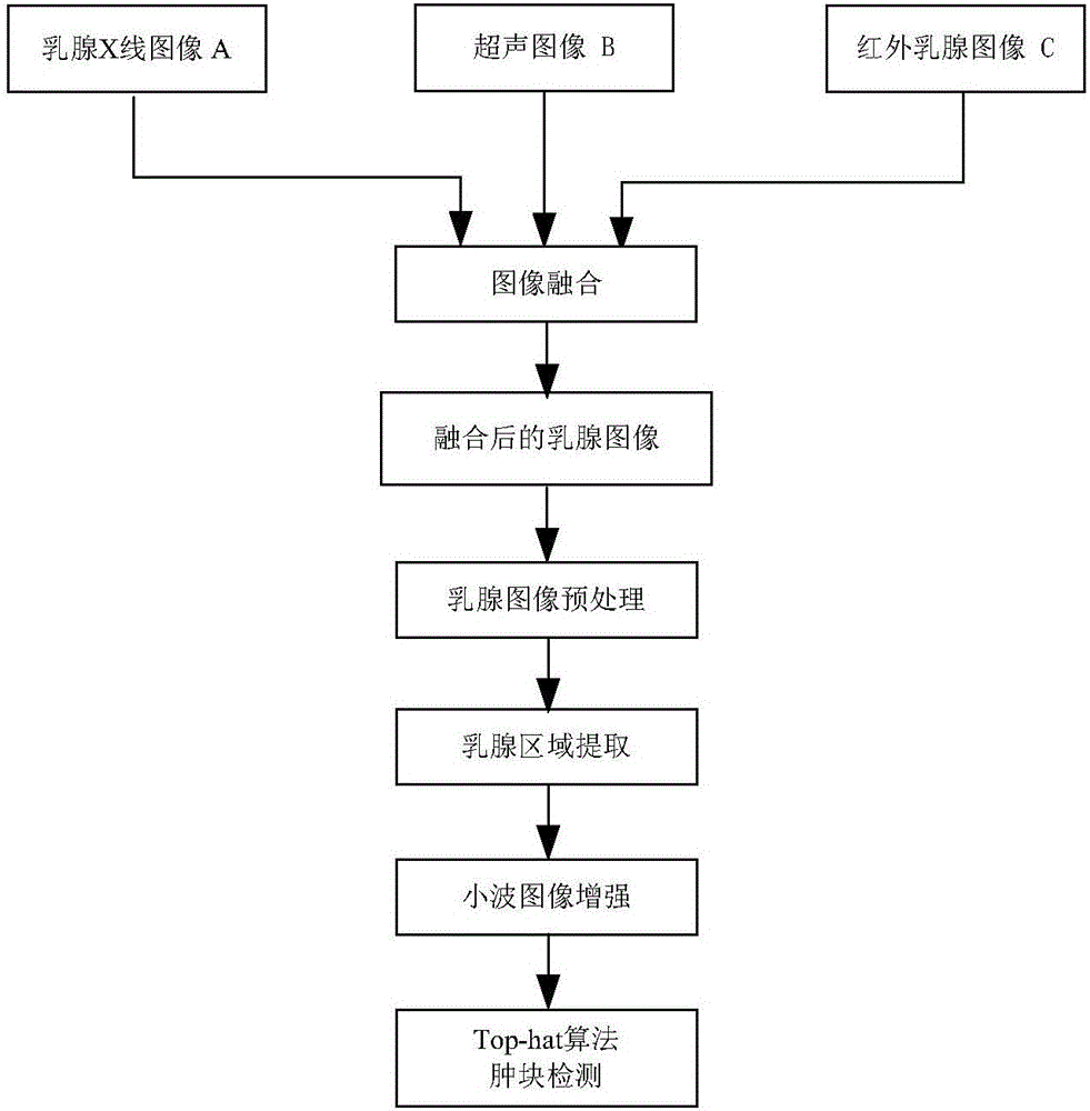
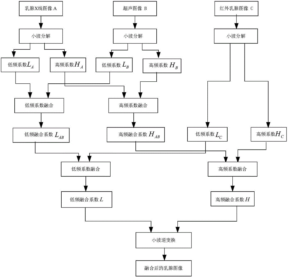
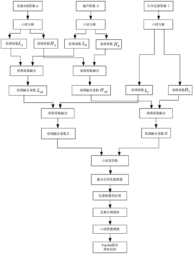
[0038] The mammary gland mass image detection method based on X-ray, ultrasound, infrared three types of image fusion is the same as embodiment 1, wherein step (1) obtains X-ray, ultrasound, infrared three types of fusion images, see figure 2 , including the following steps:

[0039] (1a) Decompose the mammography image and the ultrasound image by wavelet respectively to obtain the low-frequency coefficients and high-frequency coefficients of the mammography image and the ultrasound image respectively. Fusion, and then fuse the high-frequency coefficients of the two images after wavelet decomposition according to the high-frequency coefficient fusion rules to obtain the low-frequency coefficients and high-frequency coefficients after the fusion of the X-ray image and the ultrasonic image.

[0040] (1b) Carry out wavelet transformation to the infrared breast image to obtain its low-frequency coefficient and high-frequency coefficient. First, the low-frequency coefficient of th...

Embodiment 3

[0043] The mammary gland mass image detection method based on X-ray, ultrasonic, infrared image fusion of three types is the same as embodiment 1-2, wherein step (3) adopts grid coverage method to segment the mammary gland area in the three types of fusion images, including: Follow the steps below:

[0044] (3a) For the three types of fusion images after denoising, obtain the gray histogram of the breast image after denoising. The gray value of the breast image is mostly concentrated in 50-255, and the gray value less than 50 is mostly zero or close to At zero, that is, the background area, therefore, a threshold of 50 is used to binarize the image.

[0045] (3b) Use the grid covering method to segment the breast area in the three types of fused images, starting from the upper left corner of the binarized breast image area, from left to right, and from top to bottom, covering with a square area of ​​128×128 pixels In addition, every two coverage areas need to overlap with an ...


PUM


Abstract

The invention discloses an X-ray, ultrasonic and infrared image fusion-based breast mass image detection method, and mainly solves the technical problems of simplex information source and inaccurate information due to a single breast mass image. The method is implemented by the steps of fusing a breast X-ray image, an ultrasonic image and an infrared breast image; preprocessing and denoising a fused breast image; segmenting out a breast region for three types of fused images of the denoised breast image by using a square grid coverage method; performing wavelet image enhancement on the breast image from which the breast region is segmented out, and remarkably distinguishing a mass from a background region; and detecting the mass in the breast region based on a Top-hat morphological method to assist in early diagnosis of doctors. According to the method, multiple breast examination images are fused, so that information richness is improved and more accurate diagnosis information is obtained; and the image is subjected to analytic processing to detect out the mass, so that early discovery and diagnosis treatment of breast cancers are facilitated and an image basis is provided for the early diagnosis of the doctors.

Description

technical field [0001] The invention belongs to the technical field of image processing, and further relates to image fusion processing, in particular to an X-ray, ultrasonic, and infrared image fusion breast mass image detection method, which can be used for image analysis of early diagnosis of breast cancer. Background technique [0002] Breast cancer is a common malignant tumor that seriously threatens the health and even life of women all over the world. According to relevant information, since the late 1970s, the incidence of breast cancer has always been the first among female tumors, and it is increasing at a rate of about 2% every year in the world. 1 in 8 women in the U.S. will develop breast cancer in her lifetime. China is not a country with a high incidence of breast cancer, but in recent years, the growth rate of the incidence of breast cancer in my country is 1 to 2 percentage points higher than that of countries with a high incidence. According to the 2009 b...

Claims


Application Information

Patent Timeline
IPC IPC(8): G06T7/00G06T7/11G06T7/136
CPCG06T7/0012G06T2207/10048G06T2207/10116G06T2207/10132G06T2207/20064G06T2207/20221G06T2207/30068G06T2207/30096
Inventor 那彦李雪陈建春
Owner XIDIAN UNIV
Why Eureka
Try Eureka