Method for trapping single cells

A single-cell, target-cell technology, applied in the field of single-cell capture, can solve the problems that a single device cannot achieve high-throughput multi-size detection, low detection efficiency, and high cost, and achieve the effect of multi-size single-cell capture

Inactive Publication Date: 2017-08-04
广州高盛智造科技有限公司
View PDF3 Cites 10 Cited by
  • Summary
  • Abstract
  • Description
  • Claims
  • Application Information

AI Technical Summary

Problems solved by technology

However, the Cell-Search detection system has the problems of high cost, low detection efficiency, and the inability of a single device to achieve high-throughput multi-size detection

Method used


Image

  • Method for trapping single cells
  • Method for trapping single cells
  • Method for trapping single cells

Examples


Embodiment 1

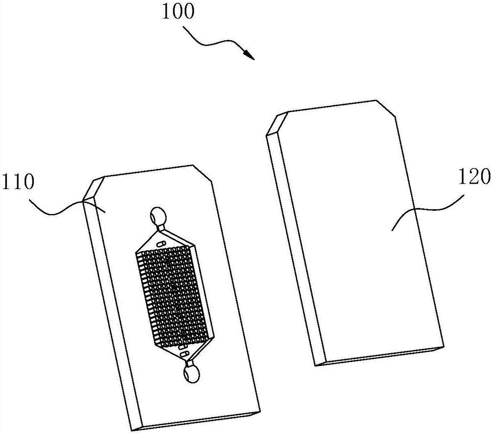
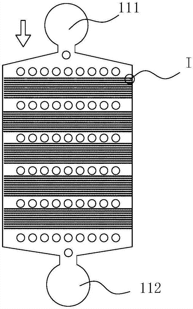
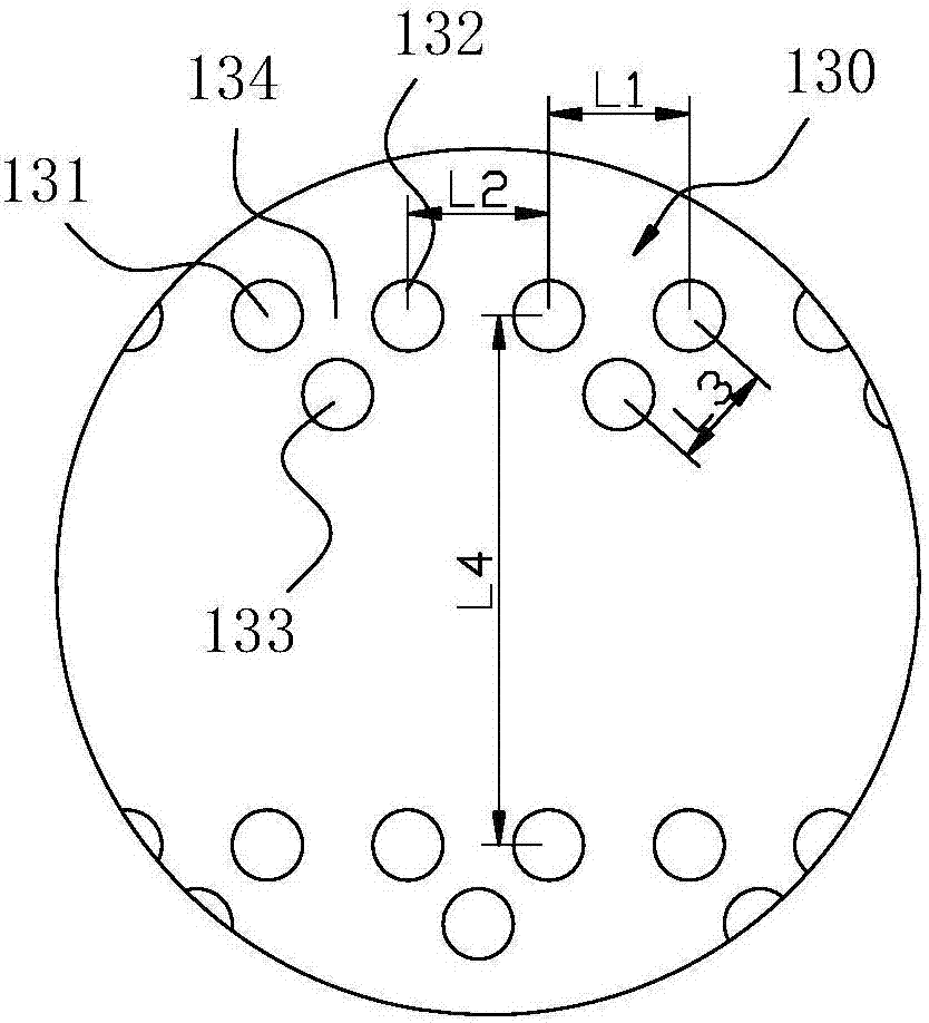
[0048] Such as Figure 1~5 As shown, in this embodiment, a single cell capture method according to the present invention provides several microfluidic chips 100, and each microfluidic chip 100 has several single cell capture units 130 of different specifications, The cell liquid containing the target cells flows through the plurality of microfluidic chips 100, and the target cells are respectively captured by different single cell capture units 130 according to different physical sizes, and the target cells not captured by the single cell capture units 130 with matching specifications Cells continue to flow to the next single cell capture unit 130 until captured or pass through all single cell capture units 130 .

[0049] In this solution, several specifications of single cell capture units 130 are set, and the structures and sizes of the single cell capture units 130 on different microfluidic chips 100 can be set to be different, so that target cells of different sizes can be...

Embodiment 2

[0065] Such as Figure 6-9 As shown, in this embodiment, a single cell capture method according to the present invention provides several microfluidic chips 200, and each microfluidic chip 200 has several single cell capture units 230 of different specifications, The cell liquid containing the target cells flows through the plurality of microfluidic chips 200, the target cells are respectively captured by different single cell capture units 230 according to different physical sizes, and the target cells not captured by single cell capture units 230 of matching specifications Cells continue to flow to the next single cell capture unit 230 until captured or pass through all single cell capture units 230 .

[0066] After the target cells are captured, the single cell capture unit 230 that has captured the target cells is positioned by means of fluorescence imaging, and the captured target cells are imaged.

[0067] The difference between this embodiment and the above embodiments...

Embodiment 3

[0076] Such as Figure 10-13As shown, in this embodiment, a single cell capture method according to the present invention provides several microfluidic chips 300, and each microfluidic chip 300 has several single cell capture units 330 of different specifications, The cell liquid containing the target cells flows through the plurality of microfluidic chips 300, and the target cells are respectively captured by different single cell capture units 330 according to different physical sizes, and the target cells not captured by the single cell capture units 330 with matching specifications Cells continue to flow to the next single cell capture unit 330 until captured or pass through all single cell capture units 330 .

[0077] After the target cells are captured, the single cell capture unit 330 that has captured the target cells is positioned by means of fluorescence imaging, and the captured target cells are imaged.

[0078] The microfluidic chip 300 includes a first structural...


PUM


Abstract

The invention discloses a method for trapping single cells. The method is implemented by the aid of a plurality of micro-fluidic chips. A plurality of single cell trapping units with different specifications are arranged on each micro-fluidic chip, cell sap with target cells can flow through the micro-fluidic chips, the target cells can be trapped by the different single cell trapping units according to physical size difference, and the target cells which are not trapped by the single cell trapping units with the matched specifications can continue flowing to the next single cell trapping units until the target cells are trapped or flow through all the single cell trapping units. According to the scheme, the method has the advantages that the single cell trapping units with the different specifications are set, the structures and the sizes of the single cell trapping units on the different micro-fluidic chips can be differently set, accordingly, the target cells with the different sizes can be trapped in the mode, and the purpose of trapping the single cells with the diversified sizes in a high-flux manner can be achieved.

Description

technical field [0001] The invention relates to the technical field of bioengineering, in particular to a method for capturing single cells. Background technique [0002] For a long time, in the clinical diagnosis and treatment of cancer, tissue cell biopsy is usually used to confirm the diagnosis and track the treatment effect. This method will not only bring physical trauma to the patient, but also be expensive. [0003] A better way to diagnose cancer is to be able to detect tumor cells free in the blood, that is, the detection of circulating tumor cells (CTCs, circulating tumor cells). Since Ashowworth reported and first proposed the concept of CTCs in 1869, CTCs have been considered an important factor in cancer metastasis, but the content of CTCs in the blood circulation system is very small, generally only 1 to 10 in 1 mL of blood (CTCs in advanced patients) number will increase). With the update of tumor biology theory and the improvement of CTCs detection technolo...

Claims


Application Information

Patent Timeline
Patent Type & Authority Applications(China)
IPC IPC(8): C12M1/00
CPCC12M47/04
Inventor 沈镝郑萍曲海洪
Owner 广州高盛智造科技有限公司
Why Eureka
Try Eureka