Pelvis CT three-dimensional reconstruction image postprocessing method based on coordinate system

A three-dimensional reconstruction and post-processing technology, applied in image data processing, image enhancement, image analysis, etc., can solve problems such as insufficient guidance for accurate reset

Active Publication Date: 2017-08-04
WEST CHINA HOSPITAL SICHUAN UNIV
View PDF5 Cites 14 Cited by
  • Summary
  • Abstract
  • Description
  • Claims
  • Application Information

AI Technical Summary

Problems solved by technology

However, it is not enough for the three-dimensional display of fractures, especially the accurate reduction guidance of unstable fractures.

Method used


Image

  • Pelvis CT three-dimensional reconstruction image postprocessing method based on coordinate system
  • Pelvis CT three-dimensional reconstruction image postprocessing method based on coordinate system
  • Pelvis CT three-dimensional reconstruction image postprocessing method based on coordinate system

Examples


Embodiment 1

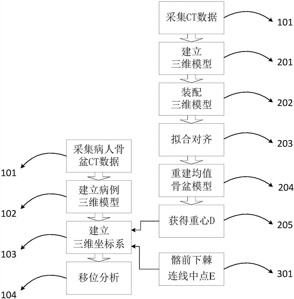
[0057] 201) collect CT data:

[0058] Screen the original data of N healthy adult pelvic CT scans on the same CT machine. The scanning parameters are: voltage 120V, slice thickness 1mm, slice distance 1mm; scan plane: align the crosshairs with the upper border of the pubic symphysis and double anterior superior iliac spines Connecting the midpoint, the lower part is to the upper part of the femur, and the upper part is located in the mid-upper abdominal plane; it is stored in DICOM format and recorded on a CD-ROM.

[0059] 202) Build a 3D model:

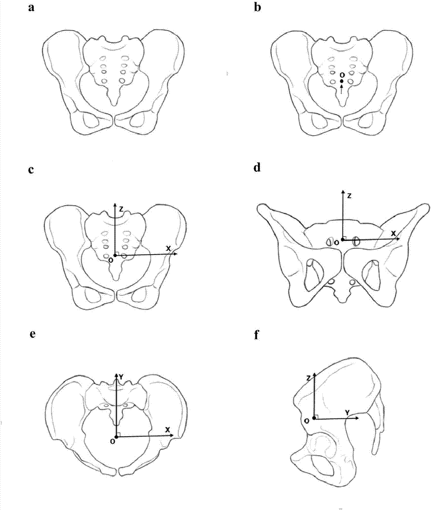
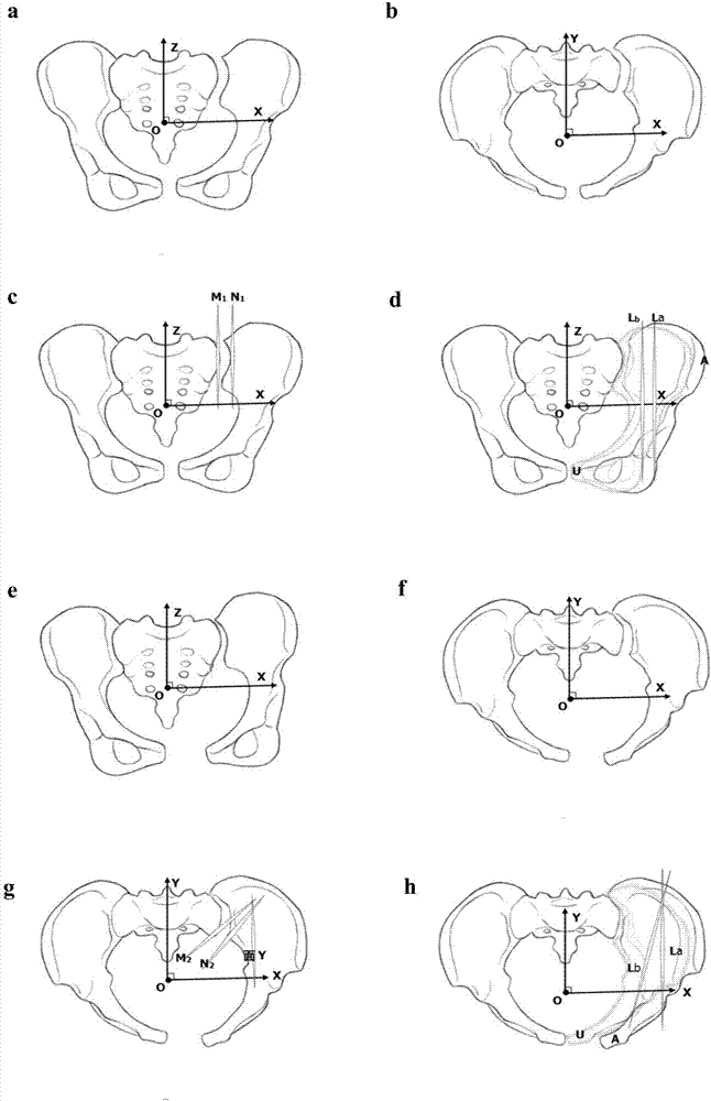
[0060] Open the 3D reconstruction software, select all the image files of the target, import the original 2D CT data by default, and set the image orientation according to the anatomical position to obtain the 3D model A i . Perform threshold segmentation according to the set bone tissue threshold (>226HU) on the menu bar, and perform region growing to remove noise from the image. Execute Edit Masks, use tools Erase and Boolean op...

Embodiment 2

[0089] Embodiment 2: Since the method of obtaining the coordinate origin D in Embodiment 1 is relatively complicated, the midpoint E of the line connecting the anterior inferior iliac spine close to point D can be used instead in clinical practice, which is especially suitable for qualitative analysis of pelvic displacement or For rough quantitative calculations, see Table 1 for specific data.

[0090] 101) Collect patient pelvic CT data:

[0091] The case data of two-dimensional CT of the pelvis before operation were collected.

[0092] 102) Establish a three-dimensional model of the case:

[0093] Using the case data, establish a three-dimensional model of the case, remove the femur and lumbar spine of the three-dimensional model of the case, and obtain the three-dimensional model of the pelvis of the case. Specifically, after the three-dimensional model of the case is established, threshold segmentation is performed, and the original data image is denoised by region growth...


PUM


Abstract

The invention discloses a pelvis CT three-dimensional reconstruction image postprocessing method based on a coordinate system and belongs to the medical image processing field. The method comprises the following steps of 101) collecting patient pelvis CT data; collecting pre-operation pelvis two-dimensional CT case data of a patient; 102) establishing a three-dimensional case model; using the case data, through three-dimensional reconstruction, acquiring a three-dimensional pelvis model of a case; 103) establishing the coordinate system in the three-dimensional pelvis model of the case; 104) acquiring a distance shift and a rotation shift of a pelvic fracture; and establishing a mathematic model in the coordinate system in the step 103), and accurately calculating distance shifts and rotation shifts of a fracture portion projected on X, Y and Z axes. By using the method, an orthopedist can conveniently, visually and accurately know a shift mode and a degree of the pelvis fracture of the patient and can accurately guide intraoperative reset for the patient.

Description

technical field [0001] The invention relates to a medical image processing method, which is an image post-processing method for three-dimensional reconstruction of pelvic CT based on a coordinate system, in particular to a displacement analysis of pelvic fractures and an accurate quantitative method for displacement degree. Background technique [0002] Pelvic fractures are often caused by high-energy injuries, and unstable pelvic fractures are often associated with various complications, such as urinary, intestinal, and neurovascular injuries, resulting in high morbidity and mortality. In recent years, with the advancement of electronic navigation equipment, internal fixation systems, and surgical techniques, the treatment of pelvic fractures has gradually shifted to minimally invasive and precise surgery. The degree of position is the key to the success of surgical reduction, reducing complications and obtaining a good prognosis. However, due to the depth and particularit...

Claims


Application Information

Patent Timeline
Patent Type & Authority Applications(China)
IPC IPC(8): G06T7/00G06T17/00
CPCG06T7/0012G06T17/00G06T2207/10081G06T2207/30008
Inventor 项舟段鑫王洪
Owner WEST CHINA HOSPITAL SICHUAN UNIV
Why Eureka
Try Eureka