
Method for labeling antibodies by colorful fluorescent granules and test paper strip prepared from antibodies

A technology for antibody preparation and test strips, which is applied in the field of colored fluorescent particle-labeled antibodies and test strips prepared using them, can solve problems such as weak signals, inconspicuous color development of test lines, and large differences between batches of quantitative detection. The effect of high sensitivity

Inactive Publication Date: 2017-09-05
江苏雷森生物科技有限公司
View PDF14 Cites 8 Cited by
  • Summary
  • Abstract
  • Description
  • Claims
  • Application Information

AI Technical Summary

Problems solved by technology

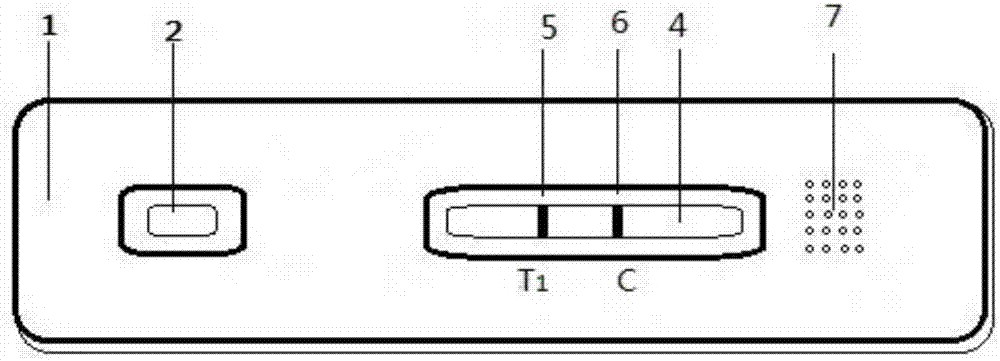
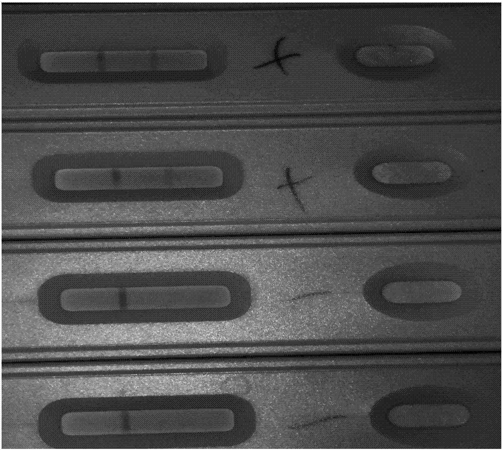
[0004] Based on this, the object of the present invention is to provide a method for labeling antibodies with colored fluorescent particles and the test strips prepared by using it to realize rapid detection of animal viruses, and to solve the problem of high cost of colloidal gold, weak signal, and unobvious color development of the test line , Quantitative detection of large differences between batches and other issues

Method used


Image

  • Method for labeling antibodies by colorful fluorescent granules and test paper strip prepared from antibodies
  • Method for labeling antibodies by colorful fluorescent granules and test paper strip prepared from antibodies

Examples


Embodiment 1

[0035] A test strip prepared by the canine distemper virus antibody labeled with colored fluorescent microspheres, comprising the following steps:

[0036] (1) Add a quantitative amount of colored fluorescent latex microsphere solution to the buffer solution for dilution, and ultrasonicate for 1.5 min to obtain a colored fluorescent microsphere dispersion.

[0037] (2) Quickly drop the activator into the dispersion of colored fluorescent latex microspheres in step (1), quickly place it on a vortex shaker and vibrate at a low speed for 1 minute, fix it on a vortex shaker, and activate it by shaking at room temperature for 30 minutes .

[0038] (3) After the activation, centrifuge the dispersion of colored fluorescent latex microspheres in step (2) at 14,000 rpm for 12 minutes at 26°C, remove the supernatant, wash with the buffer solution in step (1), and wash again After centrifugation under the same conditions, discard the supernatant and reconstitute and resuspend with the b...

Embodiment 2

[0058] A test strip prepared by a colored fluorescent microsphere-labeled feline leukemia virus antibody, comprising the following steps:

[0059] (8) Add a quantitative amount of colored fluorescent latex microsphere solution to the buffer solution for dilution, and sonicate for 1.5 min to obtain a colored fluorescent microsphere dispersion.

[0060] (9) Quickly drop the activator into the dispersion of colored fluorescent latex microspheres in step (1), quickly place it on a vortex shaker and vibrate at a low speed for 1 minute, fix it on a vortex shaker, and activate it by shaking at room temperature for 30 minutes .

[0061] (10) After the activation, the colored fluorescent latex microsphere dispersion in step (2) was centrifuged at 14000rpm at 26°C for 12min to remove the supernatant, washed with the buffer solution in step (1), and washed again with After centrifugation under the same conditions, discard the supernatant and reconstitute and resuspend with the buffer so...


PUM


Abstract

The invention provides a method for labeling antibodies by colorful fluorescent granules and a test paper strip prepared from the antibodies. Animal viruses including, but not limited to, canine distemper viruses, canine parvoviruses, canine adenoviruses, canine coronavirus, rabies viruses, feline leukemia viruses, Marek's disease viruses, Newcastle disease viruses and the like are used as targets, and the corresponding antibodies labeled by colorful fluorescent micro-spheres are prepared by the aid of chemical covalent processes and can be applied to detecting the animal viruses. The method for labeling the antibodies by the colorful fluorescent granules and the test paper strip prepared from the antibodies have the advantages that colorful detection lines with bright developed colors can appear during detection, and accordingly qualitative results can be obtained; the contents of the animal viruses in samples further can be subsequently quantitatively obtained, accordingly, test results are clear and are high in stability, and the test paper strip can be stored at the room temperature for a long time.

Description

technical field [0001] The invention relates to a method for labeling antibodies with colored fluorescent particles and a test strip prepared by using the same. The test strip prepared by this method can be used for including but not limited to canine distemper virus, canine parvovirus, canine adenovirus, canine coronavirus The rapid detection of animal viruses such as rabies virus, feline leukemia virus, Marek's disease virus and Newcastle disease virus belongs to the field of medical inspection. Background technique [0002] Compared with other microorganisms, viruses are significantly different in structure, chemical composition, mode of reproduction and susceptibility to drugs. Animal viral diseases are characterized by strong infectivity, widespread prevalence, serious harm and high incidence, so how to quickly diagnose them has become a key factor in the control and treatment of these animal viral diseases. At present, veterinary laboratories mainly use serological te...

Claims


Application Information

Patent Timeline
IPC IPC(8): G01N33/533G01N33/569
CPCG01N33/533G01N33/56983G01N2333/15G01N2333/13
Inventor 秦雷沙森朱丹丹张旭东彭雪婷陈芳
Owner 江苏雷森生物科技有限公司
Learn More