Kit for detecting keratoprotein 19 fragment and carcino-embryonic antigen contents in sample based on luminous quantum dot nanometer microemulsion probe

A technology for detecting samples and luminescent quanta, applied in the field of immunodetection, can solve the problems of difficult storage, easy to be photobleached, weak fluorescent signal, etc., and achieve the effect of short reaction time, overcoming the problem of precipitation, and strong fluorescent signal.

Inactive Publication Date: 2017-09-15
SOUTHERN MEDICAL UNIVERSITY
View PDF0 Cites 1 Cited by
  • Summary
  • Abstract
  • Description
  • Claims
  • Application Information

AI Technical Summary

Problems solved by technology

However, ordinary fluorescent substances have weak fluorescent signals, are easy to be photobleached, and are not easy to store.

Method used


Image

  • Kit for detecting keratoprotein 19 fragment and carcino-embryonic antigen contents in sample based on luminous quantum dot nanometer microemulsion probe
  • Kit for detecting keratoprotein 19 fragment and carcino-embryonic antigen contents in sample based on luminous quantum dot nanometer microemulsion probe
  • Kit for detecting keratoprotein 19 fragment and carcino-embryonic antigen contents in sample based on luminous quantum dot nanometer microemulsion probe

Examples


Embodiment 1

[0049] A kit for detecting the content of keratinin 19 fragments and carcinoembryonic antigen in a sample based on a luminescent quantum dot nanomicroemulsion probe, including an immunochromatographic test strip and a functional luminescent quantum dot nanomicroemulsion probe. Among them, the functional luminescent quantum dot nano-microemulsion probes include three types, one is a functional luminescent quantum dot nano-microemulsion probe coupled to carcinoembryonic antigen monoclonal antibody S001, and the other is a monoclonal antibody coupled to keratin 19 fragment. A functional luminescent quantum dot nano-microemulsion probe for cloning antibodies, and a functional luminescent quantum dot nano-microemulsion probe coupled to rabbit IgG.

[0050] 1. Preparation of functional quantum dot nano-microemulsion probes, the method is as follows:

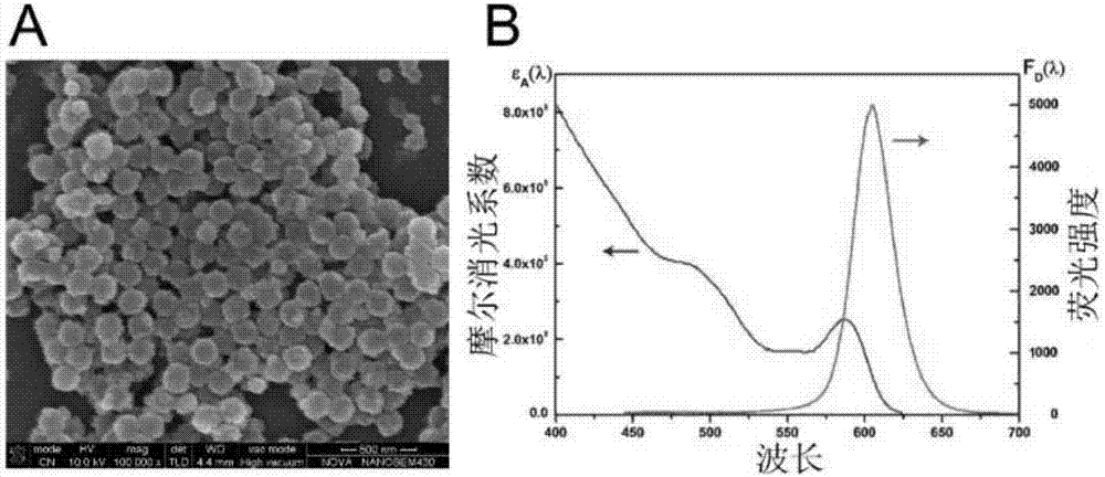
[0051] 1. Pretreatment of latex spheres: centrifuge 100µL nano latex spheres (particle size 205±3nm, mass concentration 100mg / mL, sol...

Embodiment 2

[0058] A kit for detecting the content of keratinin 19 fragments and carcinoembryonic antigen in a sample based on a luminescent quantum dot nanomicroemulsion probe, including an immunochromatographic test strip and a functional quantum dot nanomicroemulsion probe.

[0059] 1. The preparation of functional quantum dot nano-microemulsion probes is the same as in Example 1.

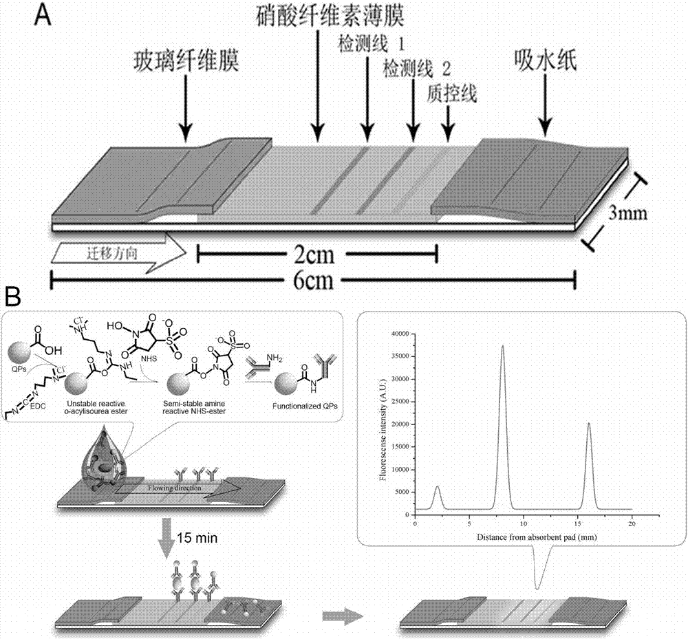
[0060] 2. The structure of the immunochromatographic test strip is as follows: figure 1 Shown, the preparation method of immunochromatographic test strip is as follows:

[0061] 1. Pretreatment of the sample pad: cut the glass fiber membrane into strips of 1.2cm×30cm, then soak the strip glass fiber membrane in the sample pad treatment solution, and soak for two hours at room temperature. After absorbing the liquid with absorbent paper, dry it overnight at 37°C, and store the dried sample pad in a dry and cool place until use.

[0062] 2. Treatment of the nitrocellulose film: Cut the nitrocellulose film i...

Embodiment 3

[0071] Methodological verification of the present invention is carried out verification to this analytical method according to routine manufacture and verification procedure in this area, and the result is as follows:

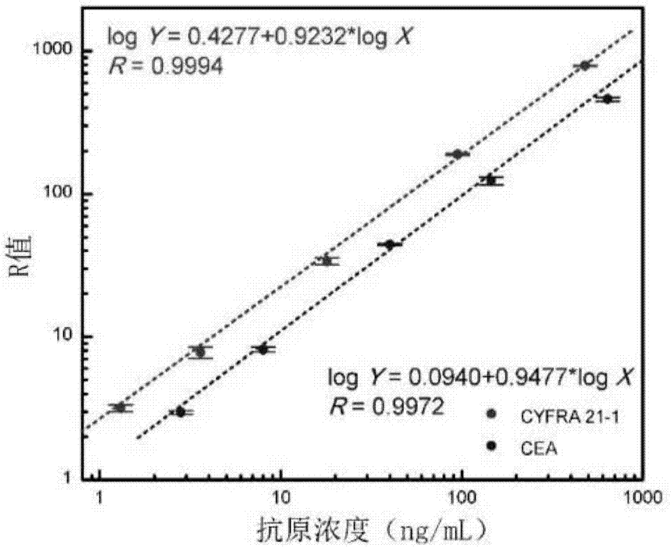
[0072] 1. Analytical sensitivity and linear range: Measure 8 times with the zero reference standard as the sample, and calculate its R average value and standard deviation. The concentration calculated by substituting the fluorescence value obtained by adding the average value of R at this point plus 2 times the standard deviation into the standard curve equation is its sensitivity. It has been determined that the sensitivity of this reagent to CYFRA 21-1 analysis is 0.16 ng / mL, and the sensitivity to CEA analysis is 0.16 ng / mL. 0.35 ng / mL. The antigens were diluted into different concentrations for determination, and the linear ranges of the measured standard curves were CYFRA 21-1: 1.3~500ng / mL; CEA: 1.2~1000ng / mL.

[0073] 2. Precision: measure the self-mad...


PUM

PropertyMeasurementUnit
concentrationaaaaaaaaaa
molecular weightaaaaaaaaaa

Abstract

The invention discloses a kit for detecting keratoprotein 19 fragment and carcino-embryonic antigen contents in a sample based on a luminous quantum dot nanometer microemulsion probe. The kit comprises an immunochromatography detection test strip and the functional quantum dot nanometer microemulsion probe. The preparation method of the functional quantum dot nanometer microemulsion probe comprises the following steps of embedding or wrapping oil soluble quantum dots with emission wavelength between 520-620nm into modified polystyrene nanoparticles to acquire quantum dot nanometer microemulsion, covalently linking the quantum dot nanometer microemulsion with a detection antibody, and performing microemulsion treatment on the surface of the microemulsion to acquire the functional quantum dot nanometer microemulsion probe. The kit provided by the invention is based on novel fluorescence immunochromatography, avoids errors caused by identification by naked eyes, and can rapidly, synchronously, quantitatively and accurately detect a plurality of biological targets.

Description

technical field [0001] The invention relates to the technical field of immune detection, in particular to a kit for detecting the content of keratin 19 fragments and carcinoembryonic antigen in a sample based on a luminescent quantum dot nano-microemulsion probe. Background technique [0002] The molecular weight of cytokeratin 19 fragment (CYFRA 21-1) is about 30KDa. Under physiological conditions, cytokeratin 19 is widely expressed on the surface of various tissue cells, commonly found in squamous epithelial and lamellar epithelial cells. In the process of malignant transformation of epithelial cells, the activated protease system accelerates the degradation of cells, and a large number of cytokeratin fragments are released into the blood circulation system. Because this soluble fragment can specifically bind to two monoclonal antibodies KS 19.1 and BM 19.21, it is called CYFRA 21-1. CYFRA21-1 is currently considered as a tumor marker mainly used to detect lung cancer. ...

Claims


Application Information

Patent Timeline
Patent Type & Authority Applications(China)
IPC IPC(8): G01N33/68
CPCG01N33/68
Inventor 刘天才吴英松陈振华
Owner SOUTHERN MEDICAL UNIVERSITY
Why Eureka
Try Eureka