Intervention type in-vivo real-time tumor imaging method and system

A tumor imaging and interventional technology, applied in the field of tumor diagnosis and research, can solve the problem that trauma cannot detect tumors in real time, and achieve the effects of accurately obtaining diagnostic information, reducing damage, and reducing pain

Active Publication Date: 2017-10-10
NORTHWEST INST OF NUCLEAR TECH
View PDF6 Cites 7 Cited by
  • Summary
  • Abstract
  • Description
  • Claims
  • Application Information

AI Technical Summary

Problems solved by technology

[0007] In order to solve the trauma caused by the biopsy process and the problem that the tumor cannot be detected in real time, the present invention provides an interventional in vivo real-time tumor imaging system and imaging method. Completable pathological diagnosis, avoiding inspection processes such as biopsy tissue staining

Method used


Image

  • Intervention type in-vivo real-time tumor imaging method and system
  • Intervention type in-vivo real-time tumor imaging method and system

Examples


Embodiment Construction

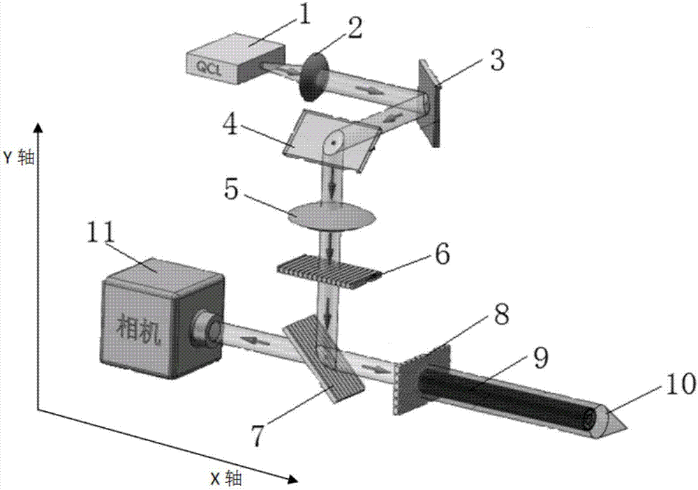
[0039] The imaging system of the invention is used to distinguish tumors and normal tissues in real time in vivo, so as to realize the purpose of diagnosing tumors. from figure 1 It can be seen that the imaging system of the present invention includes a terahertz and infrared source 1, specifically a quantum cascade laser; and a plano-convex lens 2, a first Galvano reflector 3, a second Galvano reflector 4, The lenticular lens 5, the first polarized wire grid polarizer 6, and the second polarized wire grid polarizer 7 also include a quarter-wave plate 8 and a An interventional device, and a detector 11 arranged on the transmission optical path of the second polarizing wire grid polarizer 7; the interventional device includes an arrayed fiber bundle 9 and a hollow puncture needle 10 wrapping the arrayed fiber bundle 9; the detector specifically adopts a terahertz camera.

[0040] The plano-convex lens 2 and the double-convex lens 5 converge the generated terahertz and infrared...


PUM


Abstract

Provided is an intervention type in-vivo real-time tumor imaging method and system. The imaging method comprises the steps that the polarization directions of terahertz and infrared source emergent light waves are adjusted, the light waves are conducted to the tissue to be detected through an array optical-fiber bundle, signals reflected by the tissue are converted into vertically polarized waves and then enter a detector for imaging in an incidence mode. The imaging system comprises a terahertz and infrared source, a terahertz detector, a reshaping unit, a polarization unit, a polarization conversion unit and an intervention unit. The system works in a terahertz and infrared frequency band, can perform tumor diagnosis in the human body in real time, has more advantages compared with X-ray, CT examination and other technologies, is high in spatial resolution, and has more advantages than a pathological biopsy technology in rapid, accurate and noninvasive characteristics. The intervention type in-vivo real-time tumor imaging method is safer, more accurate, quicker and more convenient and has the good application prospect in tumor diagnosis.

Description

technical field [0001] The invention belongs to the field of tumor diagnosis research, and in particular relates to an interventional in vivo real-time tumor imaging system and method. Background technique [0002] Malignant tumors are major diseases that seriously threaten human health. The incidence of malignant tumors in China has ranked first in the world, and it has become the main cause of death in my country in recent years. Continuously improving the diagnostic level of tumors is of great significance to improving the curative effect, prognosis and quality of life of cancer patients. The main methods widely used in clinical diagnosis of tumors are imaging examinations and pathological biopsy techniques, but these methods have certain limitations. [0003] The most commonly used imaging examination methods in clinical practice include X-ray, computed tomography (CT), magnetic resonance imaging (MRI), positron emission tomography (PET) and ultrasound (US). Even with ...

Claims


Application Information

Patent Timeline
Patent Type & Authority Applications(China)
IPC IPC(8): A61B5/00
CPCA61B5/0059A61B5/0084A61B5/0086
Inventor 常超崔刚强彭文毓段峰孙钧
Owner NORTHWEST INST OF NUCLEAR TECH
Why Eureka
Try Eureka