Fluorescent protein marking method for researching fusion of osteoclast

A fluorescent protein, labeling method technology, applied in the medical field, to avoid the deviation of the results, the method is intuitive, and the accuracy and repeatability are high.

Inactive Publication Date: 2017-10-10
SICHUAN UNIV
View PDF0 Cites 1 Cited by
  • Summary
  • Abstract
  • Description
  • Claims
  • Application Information

AI Technical Summary

Problems solved by technology

What's more difficult is that it can also be used to quantitatively stud...

Method used


Image

  • Fluorescent protein marking method for researching fusion of osteoclast
  • Fluorescent protein marking method for researching fusion of osteoclast
  • Fluorescent protein marking method for researching fusion of osteoclast

Examples


Embodiment 1

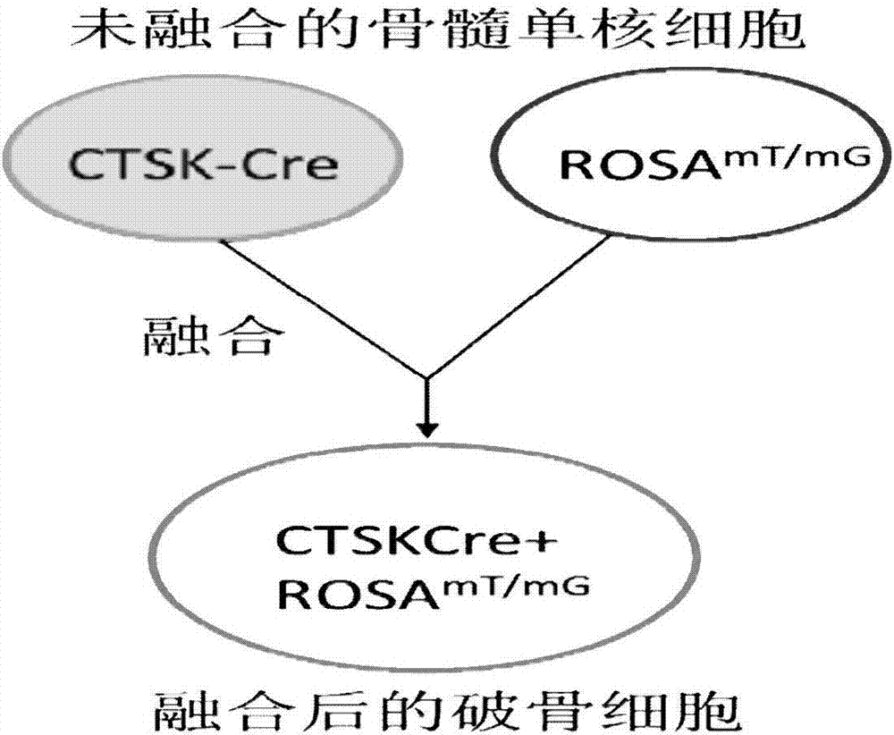
[0037] Combine CTSK-Cre transgenic mice with Rosa mTmGBone marrow mononuclear cells from transgenic mice were isolated and mixed at a ratio of 1:1.

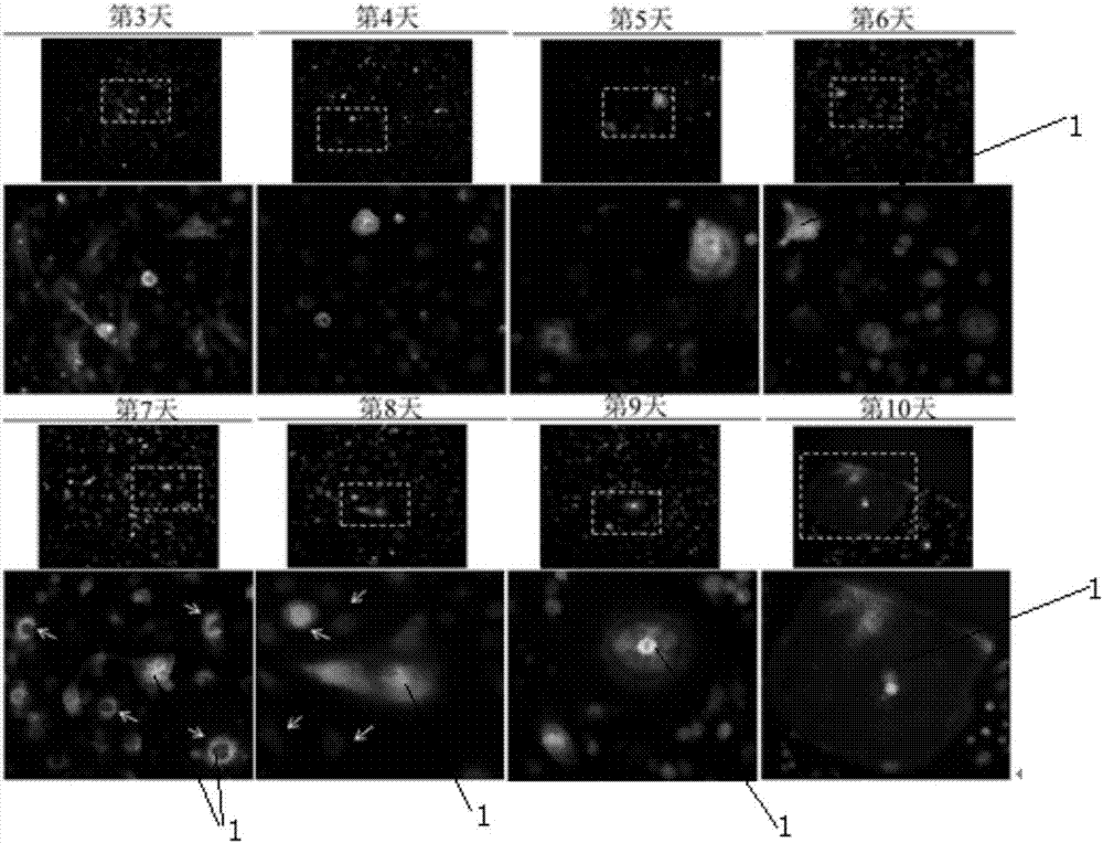
[0038] Mixed culture of CTSK-Cre transgenic mouse primary bone marrow mononuclear cells and Rosa mTmG The primary bone marrow mononuclear cells of transgenic mice were fused. Cre recombinase can specifically recognize the loxp DNA sequence, so that the gene sequence between the loxP sites is deleted or recombined, so as to achieve conditional gene targeting and finally produce green fluorescent protein.

[0039] The culture conditions are: DMEM medium (containing 10% FBS, 1% P / S), 37°C constant temperature incubator, 5% carbon dioxide, 95% oxygen.

[0040] In the culture conditions, the total amount of cells after mixing meets the following requirements: A. The total amount of cells in the 96-well plate is 3000-8000, and the volume of the medium is 100ul; The bottom area of ​​the cell is proportionally converted to obtain the ...

Embodiment 2

[0047] Combine CTSK-Cre transgenic mice with Rosa mTmG Bone marrow mononuclear cells from transgenic mice were isolated and mixed at a ratio of 1:1.

[0048] The culture conditions are: DMEM medium (containing 10% FBS, 1% P / S), 37°C constant temperature incubator, 5% carbon dioxide, 95% oxygen.

[0049] In the culture conditions, the total amount of cells after mixing meets the following requirements: A. The total amount of cells in the 96-well plate is 3000-8000, and the volume of the medium is 100ul; The bottom area of ​​the cell is proportionally converted to obtain the final mixed cell volume.

[0050] Add MSCF (final concentration: 50ng / ml) on the first day of mixed culture, and change the medium on the third day.

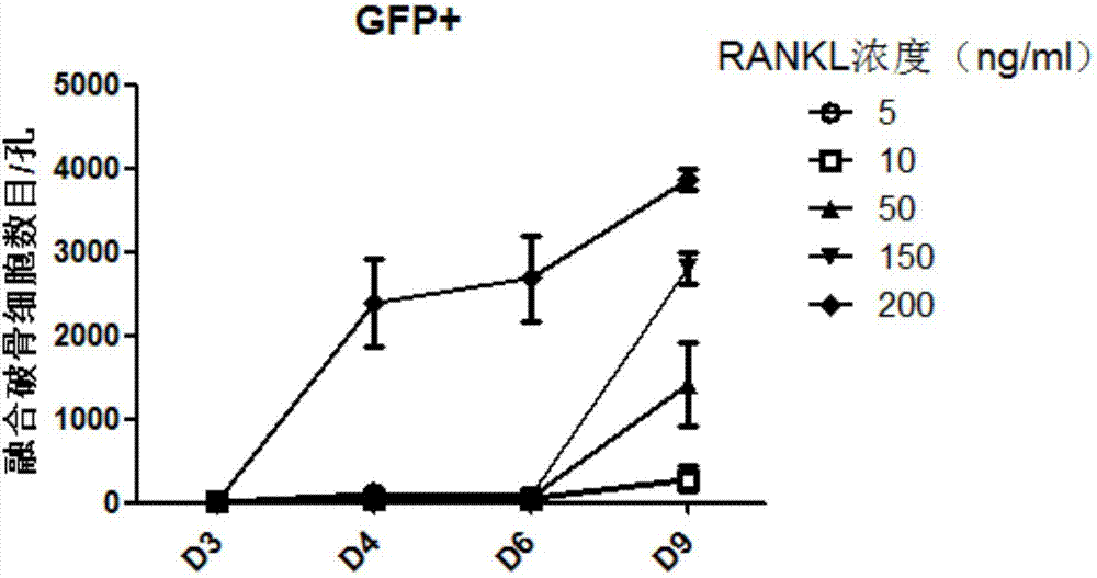
[0051] Divided into different RANKL concentration groups and different PTH concentration groups to study the effects of RANKL concentration and PTH concentration on the number and efficiency of osteoclast fusion. Add 50ng / ml MCSF to the culture medium, and ...

Embodiment 3

[0056] Other content is as embodiment 1, CTSK-Cre transgenic mouse and Rosa mTmG Bone marrow mononuclear cells from transgenic mice were isolated and mixed at a ratio of 3:1.


PUM


Abstract

The invention belongs to the field of biotechnology, and relates to a fluorescent protein labeling method for studying osteoclast fusion, which is characterized in that: primary mouse bone marrow mononuclear cells (bone marrow monocytes, BMMs) containing CTSK-Cre transgene Mix culture with RosamTmG-containing transgenic mouse primary bone marrow mononuclear cells at a ratio of 3:1-1:3, perform osteoclast differentiation induction and fluorescence microscope observation steps, the osteoclast fusion process can be visually and visually observed, thereby Observe the morphology of osteoclasts, the number of nuclei and the number of fused cells that can be quantitatively studied.

Description

technical field [0001] The invention belongs to the field of medical technology, in particular to a fluorescent protein labeling method for studying osteoclast fusion. Background technique [0002] Before this technique, there are two main types of techniques for observing osteoclast fusion: 1. live cell photography with the help of time-lapse photography microscope; 2. research with the help of the principle of virus packaging. details as follows: [0003] Time-lapse photography technology: the use of a time-lapse photography microscope to collect images of living cells in bright field at regular intervals to study whether osteoclast fusion occurs. However, even with a high magnification in bright field, it is impossible to clearly and intuitively observe the details of cell morphology and the number of nuclei during osteoclast fusion, let alone quantitatively analyze how many cells have fused. Therefore, it is impossible to study the influence of intervention factors o...

Claims


Application Information

Patent Timeline
IPC IPC(8): C12N15/06C12N5/18C12N5/10
CPCC12N5/16C12N5/0643C12N2506/115C12N2510/00
Inventor 叶玲余钒源吴凡子汪成林杨静
Owner SICHUAN UNIV
Why Eureka
Try Eureka