Polypeptide composition for detecting serum marker of autoimmune disease patient and application of polypeptide composition
A technology of serum markers and polypeptide compositions, applied in the field of medicine, can solve the problems of complex pathogenesis and unclear specific mechanisms of autoimmune diseases
- Summary
- Abstract
- Description
- Claims
- Application Information
AI Technical Summary
Problems solved by technology
Method used
Image
Examples
Embodiment 1
[0044] Example 1 Serum coating and phage adsorption and elution
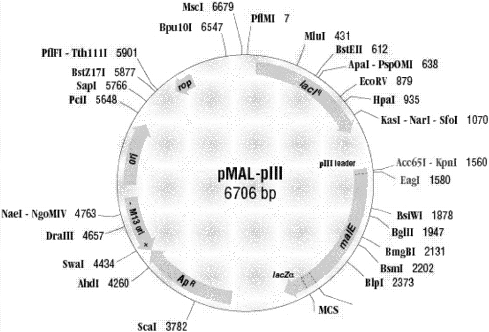
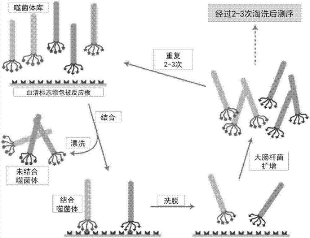
[0045] Select 30 cases of autoimmune disease patients and 30 normal human sera, mix them separately, and add them to a 96-well plate after 1:10 dilution, 150 microliters per well, incubate overnight at 4℃, 30r / min with slight shaking, then discard the wells Fill up the blocking buffer and incubate at 4°C for 2h. Pour out the blocking buffer in the well, wash it quickly with TBST (TBS+0.1v / v%Tween-20) for 6 times, and dilute 2×10 with 100μL of TBST buffer 11 The original library of pfu phage (including E.coli ER2738 host bacteria kit, produced by New England Biolabs, USA), the library plasmid map is as follows figure 1 , Add it to the wells that have been coated with normal human serum, and shake gently at room temperature for 60 minutes. Collect the phage supernatant that has not been combined with the normal human mixed serum in a sterile microcentrifuge tube. Dilute with 1 mL of TBST buffer, add to the well plat...
Embodiment 2
[0046] Example 2 Titer determination and clonal expansion of phage bound to patient mixed serum
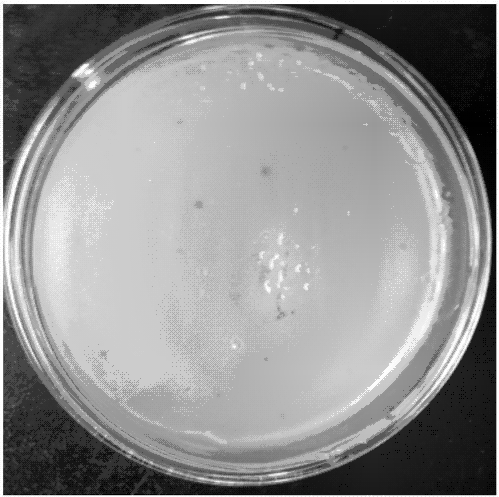
[0047] Pick the recovered single colony of E.coli ER2738 in 10ml LB medium. When the culture reaches the logarithmic growth phase, divide it into 1.5mL centrifuge tubes, each tube 200μL, and add 10μL to each tube to combine with the patient's mixed serum Incubate the phage stock solution at room temperature for 5 min, add it to the pre-warmed top agar culture tube, and immediately pour it on the LB / IPTG / Xgal plate pre-warmed at 37°C. After the plate is cooled, it is placed upside down at 37°C and incubated overnight. Check the plate and count the number of plaques on the plate with image 3 . Pick blue plaques in 1 mL of 1:100 diluted ER2738 overnight culture medium. Incubate on a shaker at 37°C at 150r / min for 4.5h. The culture was transferred to a 1.5mL centrifuge tube and centrifuged for 30s, the supernatant was transferred to a new tube, and centrifuged for another 30s. Trans...
Embodiment 3
[0048] Example 3 Determination of the ssDNA sequence of the positive phage bound to the serum of patients with autoimmune diseases
[0049] Transfer 500 μl of phage-containing supernatant to a new centrifuge tube. Add 200μl PEG / NaCl, place at room temperature for 10min, centrifuge at 10,000r / min at 4℃ for 10min, and discard the supernatant. Centrifuge for another 2 minutes and carefully remove the remaining supernatant. The pellet was completely resuspended in 100 μL of iodide buffer and 250 μL of ethanol was added. Incubate at room temperature for 10 min. Centrifuge for 10 min and discard the supernatant. The precipitate was washed with 70% ethanol and dried briefly in vacuum. The pellet was resuspended in 30μL TE [10mM Tris-HCl (pH 8.0), 1mM EDTA], and the -96gIII sequencing primer 5'-CCC TCA TAG TTA GCG TAA CG-3', 100pmol / μL, 1pmol / μL was used for automatic sequencing. The read sequence corresponds to the antisense strand of the template, write out the complementary stran...
PUM
 Abstract
Description
Claims
Application Information
 - R&D
- Intellectual Property
- Life Sciences
- Materials
- Tech Scout
- Unparalleled Data Quality
- Higher Quality Content
- 60% Fewer Hallucinations
Browse by: Latest US Patents, China's latest patents, Technical Efficacy Thesaurus, Application Domain, Technology Topic, Popular Technical Reports.
© 2025 PatSnap. All rights reserved.Legal|Privacy policy|Modern Slavery Act Transparency Statement|Sitemap|About US| Contact US: help@patsnap.com


