
Method for separating placental trophoblastic cells by using immunomagnetic bead process

A technology of trophoblast cells and immunomagnetic bead method, which is applied in the field of cell sorting of immunomagnetic bead method, can solve the problems of tediousness, low efficiency and time, and achieve the effect of high capture efficiency, convenient operation, and easy operation

Inactive Publication Date: 2017-11-03
PILOT GENE TECH HANGZHOU CO LTD
View PDF3 Cites 7 Cited by
  • Summary
  • Abstract
  • Description
  • Claims
  • Application Information

AI Technical Summary

Problems solved by technology

[0007] The object of the present invention is to provide a method for separating placental trophoblast cells by immunomagnetic bead method. The separation of trophoblast cells can be easily and quickly achieved in a few steps of incubation, and it is easy to automate, which effectively solves the problems of tediousness, inefficiency and long time in traditional methods.

Method used


Image

  • Method for separating placental trophoblastic cells by using immunomagnetic bead process
  • Method for separating placental trophoblastic cells by using immunomagnetic bead process
  • Method for separating placental trophoblastic cells by using immunomagnetic bead process

Examples


Embodiment 1

[0035] Synthesis and Coupling Efficiency Detection of HLA-G Coupling Immunomagnetic Beads

[0036] (1) Take 5mg tosyl magnetic beads (Thermo fisher M-270Epoxy (Product No. 14301)), washed twice with PBS solution (1ml each time), suspended in 100μl PBS solution.

[0037](2) Add 100 μl of HLA-G antibody and 100 μl of 3M (NH 4 ) 2 SO 4 The solution was vortexed and incubated at 37°C with tilted shaking for 24 hours to avoid the magnetic beads from settling.

[0038] (3) Wash the magnetic beads incubated in (2) 4 times with 0.1% BSA / PBS solution (1 ml each time), and suspend the magnetic beads in 1 ml 0.1% BSA / PBS solution for later use.

[0039] (4) Take 10 ul of the solution containing HLA-G antibody before and after coupling, use Quibt 3.0 Fluorometer (Life Technologies) to measure the protein content before and after the reaction, and calculate the HLA-G antibody content with successful coupling according to the difference method. The results are shown in the following t...

Embodiment 2

[0043] Culture and counting of JEG-3 cell samples

[0044] (1) JEG-3 cells were cultured and passaged in DMEM (containing 10% FBS, 1% penicillin-streptomycin double antibody solution), and JEG-3 cells were sampled during the rapid growth period for experiments;

[0045] (2) Get the JEG-3 cells in a dish (1), digest the JEG-3 cells with 0.25% trypsin digestion solution, wash the JEG-3 cells twice with PBS, collect the JEG-3 cells by centrifugation at 2500g for 5min, and dissolve the JEG-3 cells -3 Cells were suspended in 1ml PBS solution;

[0046] (3) Take 10 μl of the JEG-3 cell suspension collected in (2), dilute it 1000 times, and count the JEG-3 cells accurately with a hemocytometer. The statistics of the three counting results are shown in the table below.

[0047] JEG-3 cell count results

[0048]

Embodiment 3

[0050] Separation Efficiency Test of Immunomagnetic Beads Method

[0051] (1) Take 1.16 μl of JEG-3 cell suspension (1000 cells), add PBS solution to make up to a total volume of 100 μl;

[0052] (2) Add 200 μl of the HLA-G coupled magnetic bead suspension prepared in Example 1, and incubate at 37° C. for 2 hours with tilting shaking;

[0053] (3) Wash the magnetic bead-cell complex incubated in (2) twice with PBS solution, 0.5ml of PBS solution each time, collect the magnetic beads on the magnetic stand to remove the supernatant, add 100ul PBS solution to resuspend the magnetic beads - cell associations;

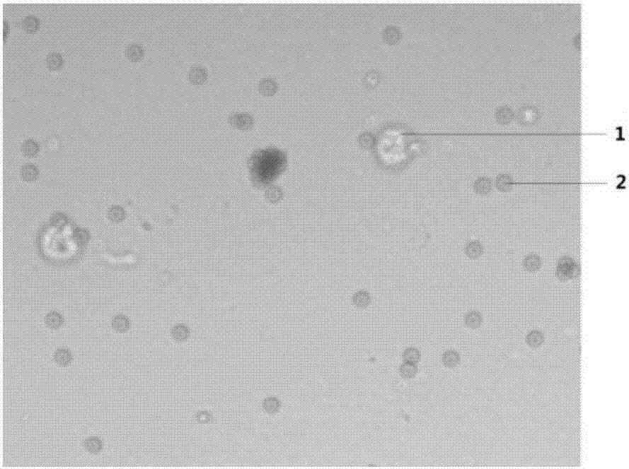
[0054] (4) Observe and distinguish the cells in the magnetic beads under a microscope, count the number of cells with a hemocytometer, the results are shown in the table below, and take pictures, the state of the magnetic bead-cell complex is as attached figure 1 Shown:

[0055] Statistical results of recovery of JEG-3 cells by immunomagnetic bead method

[0056]

[...


PUM


Abstract

The invention discloses a method for separating placental trophoblastic cells by using an immunomagnetic bead process. The method comprises the following steps: (1) preparing an immunomagnetic bead-coupled specific antibody by adding a coupling agent and an antibody capable of specifically capturing placental trophoblastic cells into immunomagnetic beads and carrying out incubation; (2) washing the antibody-coupled immunomagnetic beads obtained in the step (1) twice, and then preserving the antibody-coupled immunomagnetic beads in a preservation solution for subsequent use; (3) collecting a placental trophoblast sample and preparing a sample cell suspension by using an enzymolysis process; (4) washing the sample cell suspension obtained in the step (3) twice; (5) adding the antibody-coupled immunomagnetic beads treated in the step (2) and carrying out incubation; and (6) washing the immunomagnetic beads having undergone incubation in the step (5) twice and discarding a supernatant so as to obtain purified placental trophoblast cells. According to the invention, immunomagnetic bead cell sorting technology is employed, and the antibody-coupled immunomagnetic beads are used for specific capture of placental trophoblast cells, so trophoblastic cells can be easily and rapidly separated via a few simple steps, and automation can be easily realized.

Description

technical field [0001] The invention relates to the cell sorting technology of the immunomagnetic bead method, in particular to a method for separating placental trophoblast cells by the immunomagnetic bead method. Background technique [0002] Trophoblast cells are small individual cells that develop further after the embryo develops into a morula, and the cells begin to differentiate and expand and arrange along the inner wall of the zona pellucida, and will develop into fetal membranes and placenta in the future. Early studies have shown that placental trophoblast cells exist in the exfoliated cells of the cervix of pregnant women throughout pregnancy. Therefore, the effective isolation of trophoblast cells and the acquisition of fetal DNA are of great significance for the earliest prenatal screening and diagnosis. [0003] Many studies have shown that when the decidua and true decidua have not yet fused in early pregnancy, a certain proportion of trophoblast cells will ...

Claims


Application Information

Patent Timeline
Patent Type & Authority Applications(China)
IPC IPC(8): C12N5/073
CPCC12N5/0605C12N2509/00
Inventor 王臣罗海贝吴烈华
Owner PILOT GENE TECH HANGZHOU CO LTD
Learn More