Three-dimensional reconstruction method and device for coronary vessels, equipment and storage medium

A three-dimensional reconstruction and blood vessel technology, applied in the computer field, can solve the problems of three-dimensional reconstruction errors of coronary blood vessels, low accuracy, and relatively high requirements for known parameters.

Inactive Publication Date: 2017-11-24
SHENZHEN UNIV
View PDF2 Cites 25 Cited by
  • Summary
  • Abstract
  • Description
  • Claims
  • Application Information

AI Technical Summary

Problems solved by technology

[0004] The purpose of the present invention is to provide a method, device, equipment and storage medium for three-dimensional reconstruction of coronary vessels, aiming to solve the problem of relatively high requirements for the known degree of parameters due to the method of collecting and fusing CAG and IVUS image data in the prior art , resulting in large errors and low accuracy in the three-dimensional reconstruction of coronary vessels

Method used


Image

  • Three-dimensional reconstruction method and device for coronary vessels, equipment and storage medium
  • Three-dimensional reconstruction method and device for coronary vessels, equipment and storage medium
  • Three-dimensional reconstruction method and device for coronary vessels, equipment and storage medium

Examples


Embodiment 1

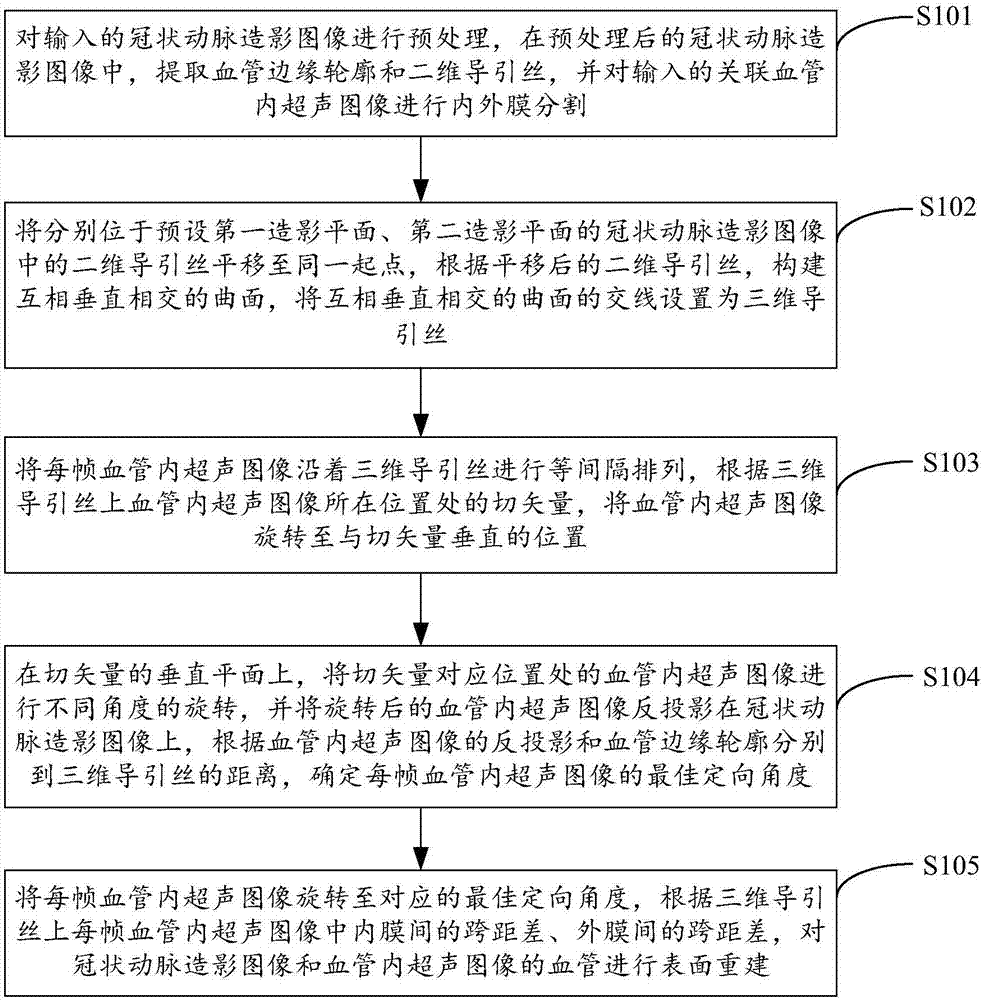
[0031] figure 1 It shows the implementation process of the three-dimensional reconstruction method of coronary vessels provided by Embodiment 1 of the present invention. For the convenience of description, only the parts related to the embodiment of the present invention are shown, and the details are as follows:

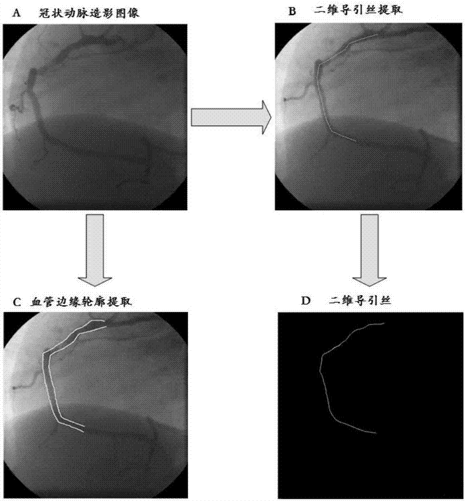
[0032] In step S101, the input coronary angiography image is preprocessed, and in the preprocessed coronary angiography image, the vessel edge contour and the two-dimensional guide wire are extracted, and the input correlative intravascular ultrasound image is subjected to intima-intima segmentation.

[0033] In the embodiment of the present invention, the input coronary angiography image and the corresponding or associated intravascular ultrasound image may come from the medical database provided by the hospital, wherein the coronary angiography device can record the patient's coronary angiography image from multiple angles , the input coronary angiography images ...

Embodiment 2

[0055] Image 6 The structure of the three-dimensional coronary vessel reconstruction device provided by the second embodiment of the present invention is shown. For the convenience of description, only the parts related to the embodiment of the present invention are shown, including:

[0056] The image processing unit 61 is configured to preprocess the input coronary angiography image, extract vessel edge contours and two-dimensional guide wires from the preprocessed coronary angiography image, and perform internal and external processing on the input associated intravascular ultrasound image. membrane segmentation;

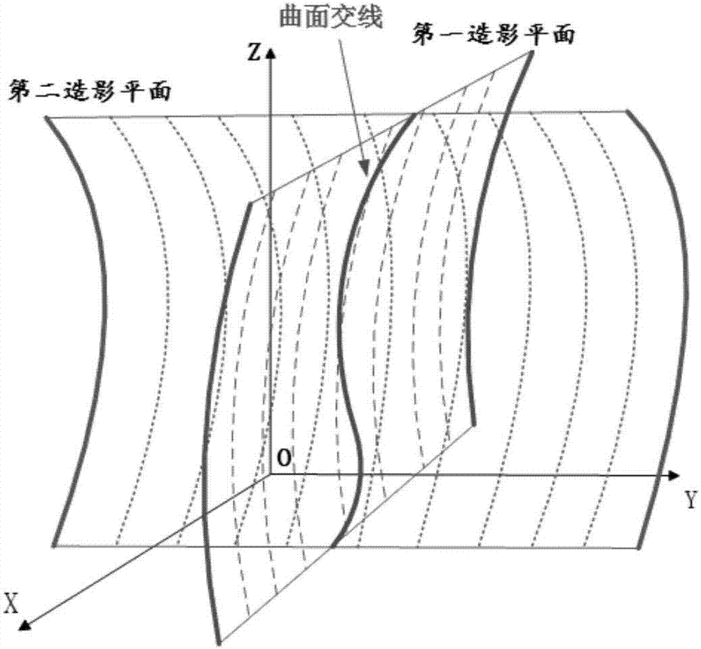
[0057] The guidewire reconstruction unit 62 is configured to translate the two-dimensional guidewires respectively located in the coronary angiography images of the preset first angiography plane and the second angiography plane to the same starting point, and according to the translated two-dimensional guidewires, Construct mutually perpendicularly intersectin...

Embodiment 3

[0070] Figure 8 The structure of the medical device provided by the third embodiment of the present invention is shown, and for the convenience of description, only the parts related to the embodiment of the present invention are shown.

[0071] The medical device 8 of the embodiment of the present invention includes a processor 80 , a memory 81 and a computer program 82 stored in the memory 81 and operable on the processor 80 . When the processor 80 executes the computer program 82, the steps in the above-mentioned method embodiments are realized, for example figure 1 Steps S101 to S105 are shown. Alternatively, when the processor 80 executes the computer program 82, the functions of the units in the above-mentioned device embodiments are realized, for example Image 6 Function of units 61 to 65 shown.

[0072] In the embodiment of the present invention, preprocessing, vessel edge contour extraction, and two-dimensional guide wire extraction are performed on coronary angi...


PUM


Abstract

The invention, which is applicable to the field of the computer technology, provides a three-dimensional reconstruction method and device for coronary vessels, equipment and a storage medium. The method comprises: carrying out pretreatment and extraction of a blood vessel edge contour and two-dimensional guide wires on a coronary artery angiography (CAG) image and carrying out inner and outer membrane segmentation on an IVUS image; carrying out translation of two-dimensional guide wires in the CAG image on a first radiography plane and a second radiography plane to a same starting point, constructing a vertical intersecting surface, and setting intersecting lines as three-dimensional guide wired; arranging each frame of IVUS image on the three-dimensional guide wire at equal interval and rotating the each frame of IVUS image to be perpendicular to a tangent vector at a corresponding position; and rotating the IVUS image on the tangent vector vertical plane and carrying out back projection to the CAG image, obtaining distances between the three-dimensional guide wires based on the back projection and the blood vessel edge contour, determining an optimal orientation angle, and then rebuilding a blood vessel surface. Therefore, the external morphological structure of the blood vessel and the inner cavity lesion information are checked simultaneously; and the efficiency and accuracy of three-dimensional reconstruction of the coronary vessel are improved.

Description

technical field [0001] The invention belongs to the technical field of computers, and in particular relates to a method, device, equipment and storage medium for three-dimensional reconstruction of coronary vessels. Background technique [0002] In recent years, the morbidity and mortality of coronary heart disease are on the rise. The main clinical diagnostic methods for coronary heart disease are coronary angiography (Coronary Angiography, CAG) and intravascular ultrasound (Intravascular UItrasound, IVUS). CAG is currently the "gold standard" for the diagnosis of coronary heart disease. CAG can be used to determine the presence of coronary artery stenosis and the location, degree, and scope of the stenosis. IVUS can be used to obtain the shape of the coronary artery wall and the degree of stenosis. However, CAG images cannot provide the structural information of the vessel wall and the extent of lesions, and IVUS cannot provide the axial position and spatial direction of t...

Claims


Application Information

Patent Timeline
Patent Type & Authority Applications(China)
IPC IPC(8): G06T17/00G06T7/149G06T7/13
CPCG06T7/13G06T7/149G06T17/00G06T2211/404A61B8/0883A61B8/0891A61B8/12A61B8/5223A61B8/483G06T2207/30101G06T2207/30021G06T2207/10116G06T2207/10132G06T7/55G06T2207/30048G16H50/30G06T7/12A61B6/5247
Inventor 张勇吴显淞张贺晔高智凡刘欣
Owner SHENZHEN UNIV
Why Eureka
Try Eureka