3D folding paper base microfluid fluorescence detection device for simultaneously detecting multiple tumor markers

A tumor marker and fluorescence detection technology, applied in the field of detection devices, can solve the problems of complex reaction procedures and unsuitability for rapid detection anytime and anywhere, and achieve the effects of simplified operation steps, simple and convenient visual reading, and high sensitivity

Active Publication Date: 2017-12-15
NANJING UNIV OF TECH
View PDF11 Cites 10 Cited by
  • Summary
  • Abstract
  • Description
  • Claims
  • Application Information

AI Technical Summary

Problems solved by technology

However, most of these methods are enzyme-linked immunosorbent immunoassays. Substrates and stop solutions need to be added to the enzyme-linked immunosorbent reaction, which makes the reaction procedu...

Method used


Image

  • 3D folding paper base microfluid fluorescence detection device for simultaneously detecting multiple tumor markers
  • 3D folding paper base microfluid fluorescence detection device for simultaneously detecting multiple tumor markers
  • 3D folding paper base microfluid fluorescence detection device for simultaneously detecting multiple tumor markers

Examples


Embodiment 1

[0034] Carbohydrate antigen 724 (CA724), a tumor marker, was detected by a 3D folded paper-based microfluidic detection device.

[0035] The specific steps of this embodiment include:

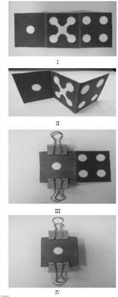
[0036] (1) Design a 3D paper-based pattern on the computer and print it with wax spray.

[0037] (2) Put the paper into the oven and bake at 150 degrees for 150 seconds and take it out.

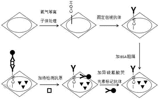
[0038] (3) After the paper base is cooled to room temperature, it is folded into a three-dimensional shape, and the third layer is treated with oxygen surface plasma for 4 minutes to modify the surface of the paper with aldehyde groups.

[0039] (4) Labeling the labeled antibody corresponding to the CA724 tumor marker with fluorescein isothiocyanate.

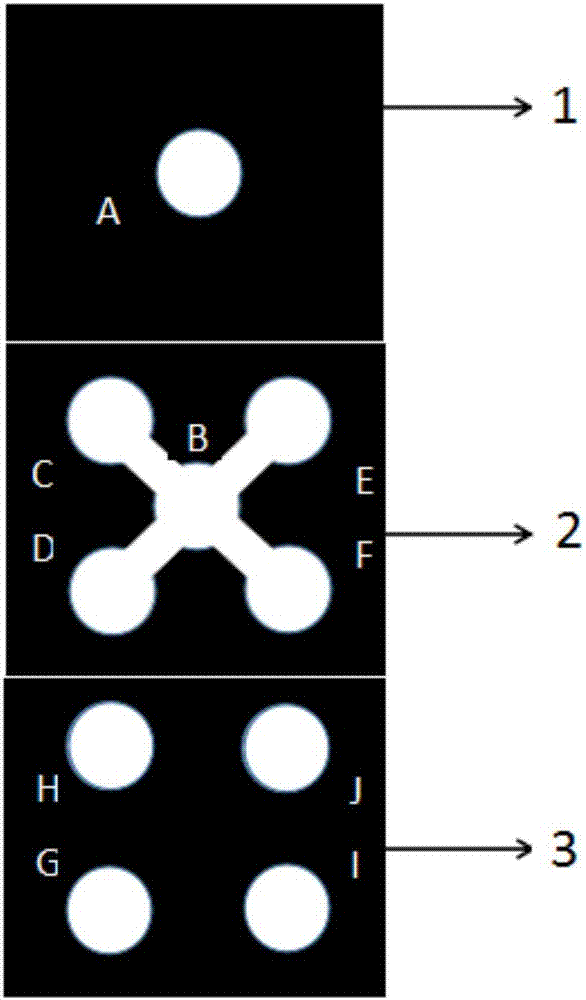
[0040] (5) Immobilize 4ul, 40ug / ml of the coated antibody corresponding to the CA724 tumor marker on the third layer of paper base G. G, I, and J are used as blank controls. After reacting at room temperature for 30min, use 20ul, 0.01molPH=7.4 Wash with phospha...

Embodiment 2

[0047] The 3D folded paper-based microfluidic detection device simultaneously detects two tumor markers, carbohydrate antigen 125 (CA125) and carbohydrate antigen 153 (CA153).

[0048] The specific steps of this embodiment include:

[0049] (1) Design a 3D paper-based pattern on the computer and print it with wax spray.

[0050] (2) Put the paper into the oven and bake at 150 degrees for 150 seconds and take it out.

[0051] (3) After the paper base is cooled to room temperature, it is folded into a three-dimensional shape, and the third layer is treated with oxygen surface plasma for 4 minutes to modify the surface of the paper with aldehyde groups.

[0052] (4) Labeling the labeled antibodies corresponding to the two tumor markers CA125 and CA153 with fluorescein isothiocyanate.

[0053] (5) 4ul and 40ug / ml of coating antibodies corresponding to the two tumor markers, CA125 and CA153, were respectively immobilized on the third layer of paper bases G and H. I and J were use...

Embodiment 3

[0060] The 3D folded paper-based microfluidic detection device simultaneously detects three tumor markers, carcinoembryonic antigen (CEA), alpha-fetoprotein (AFP), and carbohydrate antigen 199 (CA199).

[0061] The specific steps of this embodiment include:

[0062] (1) Design a 3D paper-based pattern on the computer and print it with wax spray.

[0063] (2) Put the paper into the oven and bake at 150 degrees for 150 seconds and take it out.

[0064] (3) After the paper base is cooled to room temperature, it is folded into a three-dimensional shape, and the third layer is treated with oxygen surface plasma for 4 minutes to modify the surface of the paper with aldehyde groups.

[0065] (4) The labeled antibodies corresponding to the three tumor markers CEA, AFP, and CA199 were labeled with fluorescein isothiocyanate.

[0066] (5) Immobilize 4ul, 40ug / ml of CEA, AFP, CA199 coated antibodies corresponding to the three tumor markers on the third layer of paper bases G, H, and I ...


PUM


Abstract

The invention relates to a 3D folding paper base microfluid fluorescence detection device for simultaneously detecting multiple tumor markers, belonging to the technical field of biological detection. The detection device can be used for simultaneously detecting multiple tumor markers. A method of the invention comprises the following steps: (1) producing a paper base microfluid chip; (2) preprocessing the paper base microfluid chip; (3) carrying out immunoreaction program to simultaneously detect a three-dimensional paper base; and (4) carrying out fluorescence analysis and detection. A paper base microfluid analysis platform and a fluorescence detection manner are combined together and are used for simultaneously detecting multiple tumor markers. The detection device is simple, cheap, portable, disposable and easy to use, and a beneficial platform is provided for developing countries, resource-limited regions and remote regions.

Description

technical field [0001] The invention relates to a detection device, in particular to a 3D folding paper-based microfluidic fluorescence detection device capable of simultaneously detecting multiple tumor markers, belonging to the technical field of biological detection. Background technique [0002] According to the "World Cancer Report" released by the World Health Organization and the International Agency for Research on Cancer (IARC), cancer is the leading cause of death worldwide. Early detection of multiple tumor markers facilitates cancer diagnosis and treatment monitoring, and significantly improves treatment efficiency and survival rate. However, detection of a single tumor marker is often not sufficient for an accurate diagnosis of cancer because most markers are not specific for a particular tumor. In order to improve the accuracy of cancer diagnosis, it is necessary to combine the detection of multiple tumor markers, because it can significantly improve the diagn...

Claims


Application Information

Patent Timeline
IPC IPC(8): G01N21/64G01N33/533G01N33/574
Inventor 于海东焦钰翠李林张承武刘志鹏刘金华黄维欧阳启然赖琼宇郭雪英宗丽君朱成显
Owner NANJING UNIV OF TECH
Why Eureka
Try Eureka