
Alpha fetoprotein detection kit and detection method thereof

A technology for detecting kits and alpha-fetoprotein, which is used in measurement devices, instruments, scientific instruments, etc.

Inactive Publication Date: 2018-04-10
CHANGCHUN INST OF APPLIED CHEMISTRY - CHINESE ACAD OF SCI
View PDF1 Cites 4 Cited by
  • Summary
  • Abstract
  • Description
  • Claims
  • Application Information

AI Technical Summary

Problems solved by technology

There is no report on the application of tyrosinase in the detection of alpha-fetoprotein in the prior art

Method used


Image

  • Alpha fetoprotein detection kit and detection method thereof
  • Alpha fetoprotein detection kit and detection method thereof
  • Alpha fetoprotein detection kit and detection method thereof

Examples


Embodiment 1

[0027] Preparation of tyrosinase-streptavidin conjugate

[0028] Dissolve 1 mg of tyrosinase and 0.185 mg of biotin 3-sulfo-N-hydroxysuccinimide sodium salt (molar ratio = 1:50) in 500 μL of 10 mM phosphate buffer solution (pH = 7.4), and incubate at room temperature The biotin-labeled tyrosinase was obtained in 60 min, purified with an Amicon Ultra centrifuge tube with a molecular weight cut-off of 30 kDa, centrifuged at a speed of 10,000 r / min, and centrifuged for 10 min, and repeated 3 times. Then 500 μL streptavidin (500 μg / mL) was added to the purified biotin-tyrosinase conjugate, and after incubation at room temperature for 30 min, the tyrosinase-streptavidin conjugate was obtained, and put 4°C refrigerator for later use.

Embodiment 2

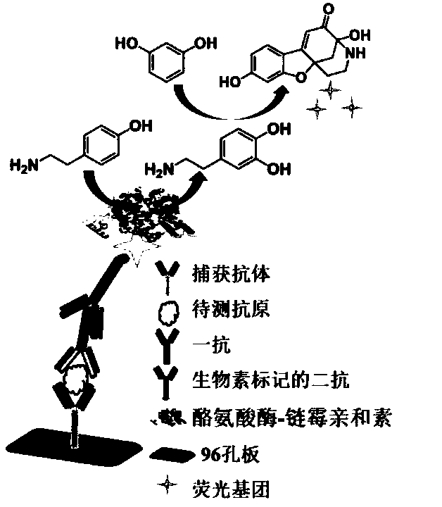
[0030] Sandwich ELISA operation steps based on tyrosinase-induced fluorescent reaction:

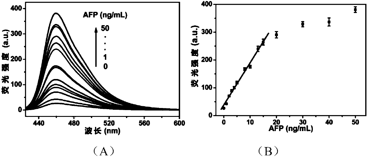
[0031] First, the mouse monoclonal antibody (anti-AFP) was diluted 200 times with coating diluent, 100 μL was added to a 96-well plate, and then left at 4° C. for overnight coating. Discard the liquid in the well and wash with 300 μL of washing buffer, add 200 μL of bovine serum albumin (0.01 mg / mL) to the well, block for 1 h at 37 °C, discard the liquid in the well, wash with 300 μL of washing buffer, and then add 100 μL of different Concentration AFP standard solution (0, 1, 2, 3, 4, 5, 7, 10, 15, 20, 30, 40, 50ng / mL) and incubate at 37°C for 1h, discard the liquid in the well and wash with 300μL of buffer wash the 96-well plate. Add 100 μL of diluted rabbit polyclonal antibody (anti-AFP, 1:500), incubate at 37°C for 1 hour, discard the liquid in the well and rinse. Add 100 μL of biotin-labeled secondary antibody (1:2000), incubate at 37°C for 1 h, discard the liquid in the well and r...

Embodiment 3

[0035] Detection of AFP content in serum of patients with liver cancer and normal people:

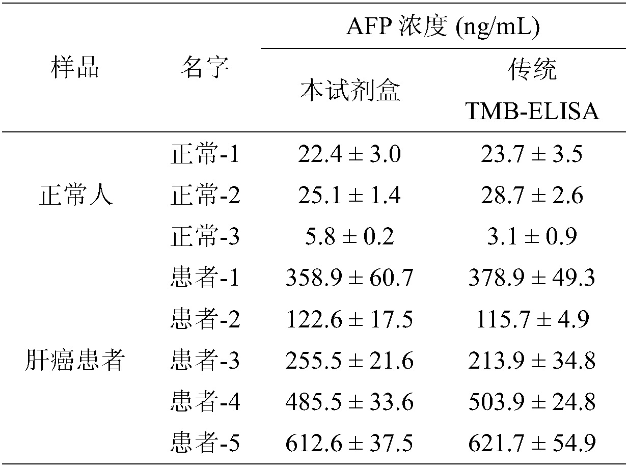
[0036] The clinical serum samples of patients with liver cancer and normal subjects were provided by the Second Hospital of Jilin University. The serum fluorescence sandwich ELISA method was basically the same as the antigen determination method in Example 2, except that the actual serum samples were used instead of the antigen standard solution. All serum samples were diluted 20-100 times before testing and added to the wells. At the same time, the traditional TMB-ELISA method was used as a control, and the AFP content in the serum was detected according to the operation steps in the TMB-ELISA-AFP kit.

[0037] As shown in Table 1, the detection results of the AFP content in the serum of normal people and patients with liver cancer show that the detection results of this kit are equivalent to those of the traditional TMB-ELISA-AFP kit, showing that the kit of the present invention can ...


PUM


Abstract

The invention provides an alpha fetoprotein detection kit and a detection method thereof, and belongs to the field of fluoroimmunoassay. The alpha fetoprotein detection kit is of a nearly-sandwiched structure formed by adsorbing a capturing antibody, AFP and a primary antibody on a 96-well plate in sequence, afterwards a secondary antibody and tyrosinase-streptavidin conjugate which are labeled bybiotin are added in sequence, conjugate of tyrosinase and the secondary antibody is specifically combined with the detecting primary antibody, finally substrate tyramine is added, dopamine is generated under the effect of tyrosinase, and is reacted with resorcinol which is added afterwards under an alkaline condition to generate perssads for strong fluorescence emission, and then quantitative detection of the AFP is achieved. The detection result of the kit is consistent with a standard ELISA method, it is proved that the kit can sensitively detect the AFP through fluorescence activation, andfinally the AFP is successfully used for diagnosing liver cancer diseases. Besides, the detection method has universality, and can be applied to detecting other antigens by simply replacing corresponding capturing antibodies and detecting primary antibodies according to different drone antigens.

Description

technical field [0001] The invention belongs to the field of fluorescence immunoassay, and in particular relates to an alpha-fetoprotein detection kit and a detection method thereof. Background technique [0002] According to recent data from the Chinese Anti-Cancer Association, nearly 6,000 people die of cancer every day in China, and cancer has become the most common cause of death. Globally, cancer will become one of the main causes of morbidity and death in the next few decades. Therefore, sensitive detection of cancer biomarkers is increasingly important for early clinical diagnosis, disease prevention and biomedical research. Methods such as electrochemistry, fluorescence, microfluidic chips, naked-eye detection, and surface plasmon resonance sensing have been successfully used to detect cancer biomarkers and have made significant progress. It is worth noting that fluorescent immunoassay has received more and more attention due to its high sensitivity, high throughput...

Claims


Application Information

Patent Timeline
IPC IPC(8): G01N33/533G01N33/558
CPCG01N33/533G01N33/558
Inventor 杨秀荣赵佳会孙健邢志财
Owner CHANGCHUN INST OF APPLIED CHEMISTRY - CHINESE ACAD OF SCI
Learn More