Fluorescent biological sensor for detecting uracil-DNA glycosylase (UDG)

A biosensor and glycosylase technology, applied in the field of biosensors, can solve the problems of long detection period, low specificity and sensitivity, and achieve the effects of short detection period, good specificity and simple preparation method

Inactive Publication Date: 2018-05-29
UNIV OF JINAN
View PDF6 Cites 14 Cited by
  • Summary
  • Abstract
  • Description
  • Claims
  • Application Information

AI Technical Summary

Problems solved by technology

[0003] In order to solve the problems of relatively low specificity and sensitivity and long detection period of the method for detecting UDG in the prior art, the present invention provides a method based on endonuclease-assisted rolling circle amplification with high specificity and sensitivity and fast detection speed. Amplified fluorescent biosensor for detecting DNA glycosylase and its application

Method used


Image

  • Fluorescent biological sensor for detecting uracil-DNA glycosylase (UDG)
  • Fluorescent biological sensor for detecting uracil-DNA glycosylase (UDG)
  • Fluorescent biological sensor for detecting uracil-DNA glycosylase (UDG)

Examples


Embodiment 1

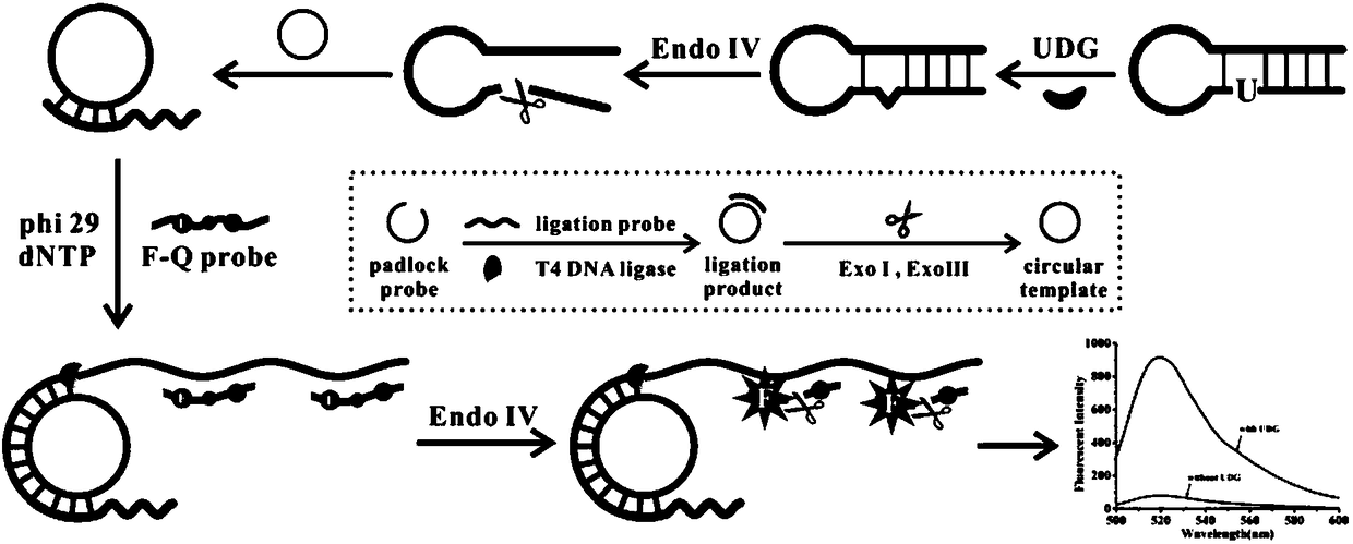
[0045] Example 1 Preparation of circular template.

[0046] Prepared with 50mM Tris-HCl, 10mM MgCl 2 , T4 DNA Ligase Reaction Buffer with 10 mM DTT and 1 mM ATP.

[0047] (1) Mix 42 μL sterilized water, 6 μL linear template (100 nM), 6 μL ligation probe (100 nM) and 6 μL 10× T4 DNA ligase buffer, denature at 95°C for 5 min, then Slowly cool down to room temperature to complete the hybridization, then add 3 μL of T4 DNA ligase (60 U / μL) to the reaction system, and react at 16°C for 20 hours; after that, the reaction system is placed in a water bath at 65°C for 15 minutes , inactivate the T4 DNA ligase in the system;

[0048] (2) Add 3 μL of exonuclease I (20 U / μL) and 3 μL of exonuclease III (100 U / μL) to the above reaction system and react at 37°C for 2 h; then place the reaction system in a water bath at 85°C Heated for 10 min to obtain a circular template, which was stored at 4°C for future use.

Embodiment 2

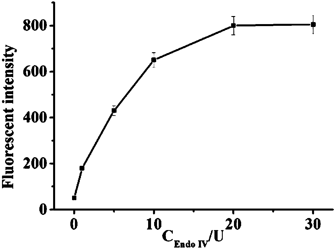
[0049] Example 2 The change of fluorescence intensity with the concentration of endonuclease IV.

[0050](1) Mix 3 μL circular template (10 nM), 3 μL hairpin probe (10 nM), 3 μL dNTP (1 mM), 2 μL phi29 DNA polymerase (1 U / μL), 2 μL endonuclease IV (The final concentrations are 1 U / mL, 5 U / mL, 10 U / mL, 20 U / mL, and 30 U / mL) in 3 μL phi29 DNA polymerase buffer, and then add 3 μL UDG enzyme solution (1 U / mL), after mixing, react at a constant temperature of 37°C for 90 min;

[0051] (2) Add 2 μL of fluorescent probe (10 μM) to the solution in step (1), mix well and react at a constant temperature of 37°C for 30 min;

[0052] (3) Take 32 μL of the solution obtained in step (2) and dilute it with water to 100 μL, and then perform fluorescence detection; set the excitation wavelength to 486 nm, the emission wavelength to 518 nm, and the detection range to 450 nm-530 nm, and read the change of the fluorescence signal .

[0053] See the test results figure 2 , it can be seen from...

Embodiment 3

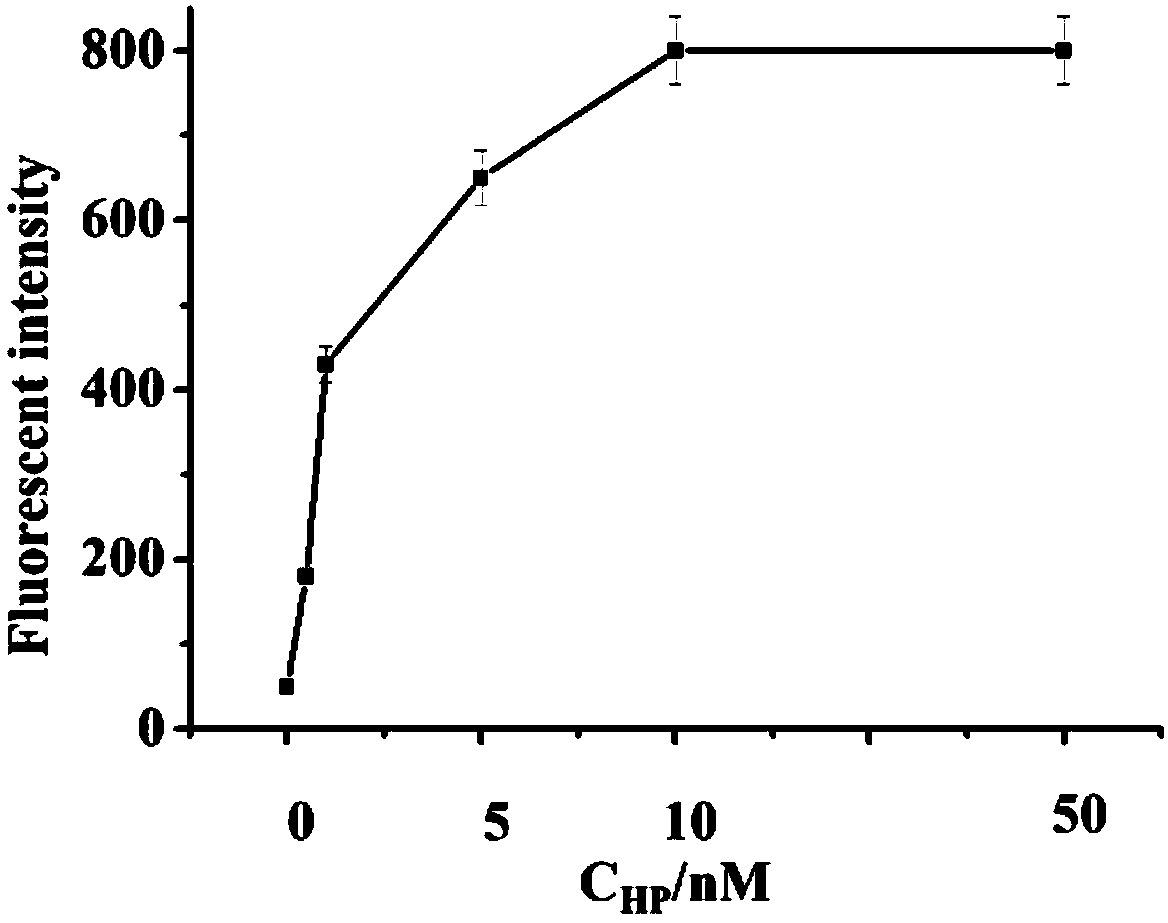
[0054] Example 3 Fluorescence intensity changes with hairpin probe concentration.

[0055] (1) Mix 3 μL circular template (10 nM), 3 μL hairpin probe (final concentrations are 0.5 nM, 1 nM, 5 nM, 10 nM, 50 nM), 3 μL dNTP (1 mM), 2 μL phi29 DNA Mix polymerase (1 U / μL), 2 μL endonuclease IV (20 U / mL) in 3 μL phi29 DNA polymerase buffer, add 3 μL UDG enzyme solution (1 U / mL), mix well Afterwards, 37°C constant temperature reaction for 90 min;

[0056] (2) Add 2 μL of fluorescent probe (10 μM) to the solution in step (1), mix well and react at a constant temperature of 37°C for 30 min;

[0057] (3) Take 32 μL of the solution obtained in step (2) and dilute it with water to 100 μL, and then perform fluorescence detection; set the excitation wavelength to 486 nm, the emission wavelength to 518 nm, and the detection range to 450 nm-530 nm, and read the change of the fluorescence signal .

[0058] See the test results image 3 , it can be seen from the figure that with the increas...


PUM


Abstract

The invention provides a fluorescent biological sensor for detecting uracil-DNA glycosylase (UDG). The fluorescent biological sensor comprises an annular template, a hairpin probe, a fluorescent probe, dNTP, phi 29 DNA polymerase and endonuclease IV. The biological sensor disclosed by the invention has the advantages of good specificity, high flexibility, moderate reaction condition and quick reaction speed; by means of silver cluster fluorescent detection, the biological sensor has the advantages of convenience in operation, short detection period, easiness in carrying, low technological cost, suitability for an industrial inexpensive requirement; a preparation method has the advantages of simpleness, stable performance and good repeatability.

Description

technical field [0001] The invention belongs to the technical field of biosensors, and relates to a fluorescent biosensor for detecting DNA glycosylase based on endonuclease amplification and rolling circle amplification. Background technique [0002] Hydrolysis of cytosine to uracil is the most common form of DNA hydrolytic damage, resulting in the conversion of G:C base pairs to A:U base pairs during DNA replication. Uracil-DNA glycosylase (UDG) is a protease with essential excision repair enzyme activity that plays a crucial role in maintaining genome integrity. As the initiator of the base excision repair pathway and a "scout" for undesired abnormal bases, UDG can act on the recognition and catalysis of single-stranded or double-stranded DNA due to its high specificity for uracil Hydrolysis and cleavage of N-glycosidic linkages on Subsequently, an apurinic and apyrimidinic site (AP site) is created and triggers the DNA repair process, which is removed by the coordinate...

Claims


Application Information

Patent Timeline
Patent Type & Authority Applications(China)
IPC IPC(8): G01N21/64
CPCG01N21/6402G01N21/6486
Inventor 黄加栋王敬锋王玉刘素
Owner UNIV OF JINAN
Why Eureka
Try Eureka