A kind of preparation method of amniotic membrane fixer

A technology of fixator and amniotic membrane, which is applied in the fields of prosthesis, ophthalmic treatment, medical science, etc., can solve the problems of inability to meet individual requirements, hard PMMA material, and difficulty in matching patients, so as to treat ocular surface damage and meet individual needs. Chemical requirements, the effect of high hydrophilic properties

Active Publication Date: 2021-03-30
广州锐澄医疗技术有限公司
View PDF4 Cites 0 Cited by
  • Summary
  • Abstract
  • Description
  • Claims
  • Application Information

AI Technical Summary

Problems solved by technology

However, due to the hard texture and fixed size of the PMMA material, it is difficult to perfectly match the patient and cannot meet individual requirements

Method used


Image

  • A kind of preparation method of amniotic membrane fixer
  • A kind of preparation method of amniotic membrane fixer
  • A kind of preparation method of amniotic membrane fixer

Examples


Embodiment 2

[0031] Embodiment 2 The mold of the amniotic membrane fixer

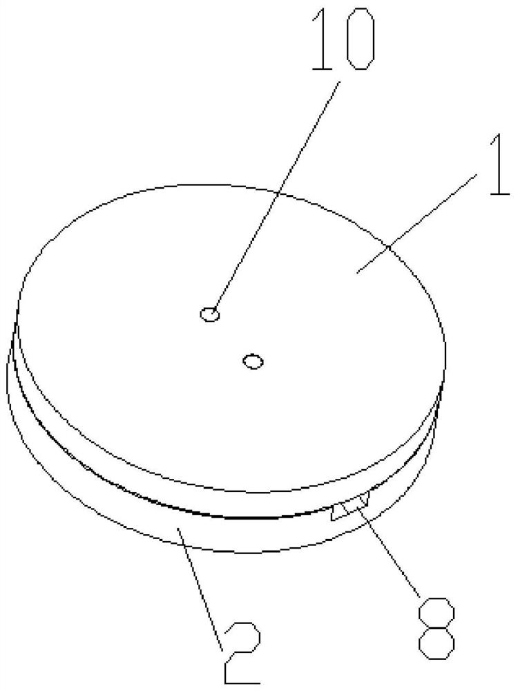
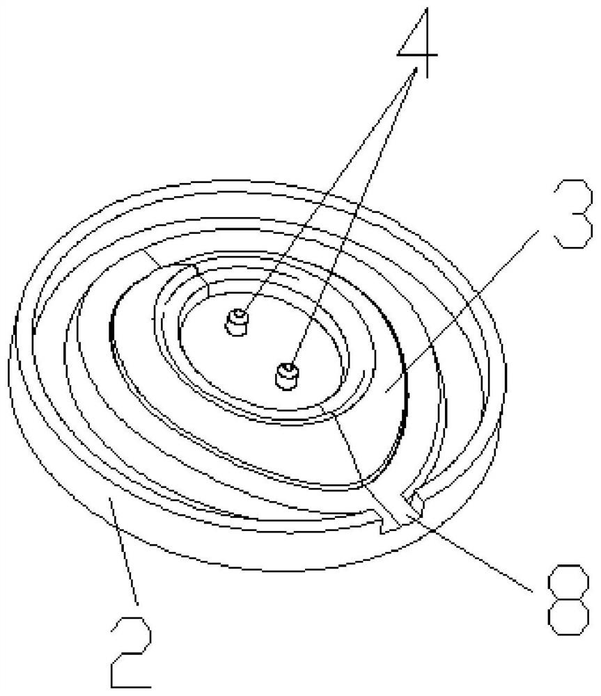
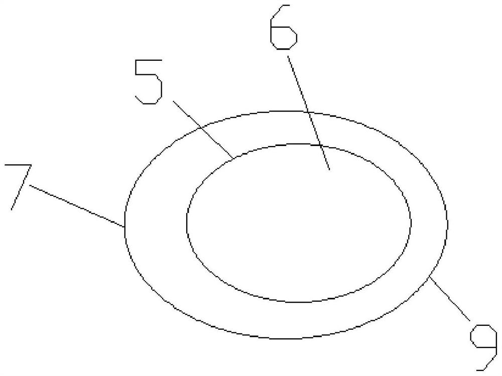
[0032] An embodiment of the mold of the amniotic membrane fixer in the present invention, as figure 1 and 2 As shown, it includes an upper mold 1 and a lower mold 2, the upper mold 1 is provided with an upper core (not shown in the figure), the lower mold 2 is provided with a lower core 3, and the lower core 3 is provided with a There is a protrusion 4, and the upper core is provided with a groove 10 that matches the protrusion 4, so that the upper mold clamping 1 and the lower clamping mold 2 can be molded more firmly, and the formed product—the size of the amniotic membrane fixer More standard and unified, after the upper clamping mold 1 and the lower clamping mold 2 are connected together, the middle parts of the upper mold core and the lower mold core 3 are tightly connected to form the amniotic membrane fixer (see image 3 ), the inner ring 5 in the middle of the mold, the amniotic membrane fixer correspondin...

Embodiment 3

[0033] The preparation method of embodiment 3 amniotic membrane fixer

[0034] A kind of embodiment of the preparation method of amniotic membrane fixer of the present invention, comprises the steps:

[0035] (1) Dissolving hydroxyethyl methacrylate in water, adding macromolecular crosslinking agent polyethylene glycol diacrylate, initiator 4-chlorobenzophenone and glycerin in sequence, vigorously shaking and mixing to obtain a mixture;

[0036] (2) Ultrasonic method is used to remove the air bubbles in the mixture obtained in step (1), and then injected into the amniotic membrane fixer mold of Example 2;

[0037] (3) Place the mold of the amniotic membrane fixer in a UV curing machine, irradiate with ultraviolet light for 5 minutes, and cure and form;

[0038] (4) Take out the molded amniotic membrane fixer from the mold, soak and wash it with clean water for 4 days, take it out, and dry it to obtain the amniotic membrane fixer. See Example 1 for its structure.

[0039] Whe...

Embodiment 4

[0040] The preparation method of embodiment 4 amniotic membrane fixer

[0041] A kind of embodiment of the preparation method of amniotic membrane fixer of the present invention, comprises the steps:

[0042] (1) Dissolve hydroxyethyl methacrylate in water, and then add the mixture of macromolecular crosslinking agent polyethylene glycol diacrylate and ethylene glycol dimethacrylate (polyethylene glycol diacrylate and dimethyl The mass ratio of ethylene glycol acrylate is 1:2), initiator benzoin dimethyl ether and glycerin, vigorously shake and mix to obtain a mixture;

[0043] (2) Ultrasonic method is used to remove the air bubbles in the mixture obtained in step (1), and then injected into the amniotic membrane fixer mold of Example 2;

[0044](3) Place the mold of the amniotic membrane fixer in a UV curing machine, irradiate with ultraviolet light for 10 minutes, and cure and form;

[0045] (4) Take out the molded amniotic membrane fixer from the mold, soak and wash it wi...


PUM

PropertyMeasurementUnit
thicknessaaaaaaaaaa
elastic modulusaaaaaaaaaa

Abstract

The invention discloses a method for preparing an amniotic membrane fixator. The method for preparing the amniotic membrane fixator includes the following steps of: (1) dissolving hydroxyethyl methacrylate in water, and sequentially adding macromolecular crosslinking agents, initiator and glycerin for shaking violently and mixing evenly to obtain a mixture; (2) removing bubbles in the mixture obtained in the step (1) to be injected into an amniotic fixator mold; (3) placing the amniotic fixator mold in a UV curing machine for curing and molding under UV irradiation for 5 to 30 minutes; (4) removing the molded amniotic fixator from the mold to be soaked and washed with water for 2 to 4 days and taken out and dried to obtain the amniotic membrane fixator. With the hydroxyethyl methacrylate (HEMA) as a base material, and a casting molding method is used to prepare the amniotic membrane fixator of an annular ocular surface, the amniotic membrane fixator has the advantages of having high hydrophilicity, excellent biocompatibility, capability of absorbing water and self expanding, and not only self-fixing effect but also special structural design, being consistent with the ocular surfacearc of patients, and satisfying personalized requirements.

Description

technical field [0001] The invention relates to an auxiliary device for ophthalmology treatment, in particular to a preparation method of an amniotic membrane fixer. Background technique [0002] In the middle and late stages of ocular surface chemical injury and thermal burn, subconjunctival scarring often leads to narrowing of the conjunctival sac, symblepharon, entropion and trichiasis, eyelid insufficiency, and even restrictive eye movement disorders. Tricky question. Repairing conjunctival defects is an important aspect of various symblephane surgeries. Conjunctiva, labial mucosa, skin grafts, and silicone sheet grafts are all used. However, the inherent defects of the graft itself and limited sources limit its application. [0003] In recent years, amniotic membrane transplantation as a biological dressing has been widely used in the treatment of ocular surface burns, and it has been confirmed that it can accelerate the epithelialization of ocular surface tissue, redu...

Claims


Application Information

Patent Timeline
Patent Type & Authority Patents(China)
IPC IPC(8): C08F283/06C08F220/20C08F222/14A61F9/00
CPCA61F9/0017A61F2210/0061A61F2220/0008C08F220/20C08F283/065C08F222/102
Inventor 李锐聪郑亚新郭少成曾晨光杨习锋
Owner 广州锐澄医疗技术有限公司
Why Eureka
Try Eureka