Preparation method of 3D (Three Dimensional) brain organ

An organoid and brain technology, applied in the field of preparing 3D cerebral organoids from human neurospheres, can solve the problems of limitations, 3D neocortical corpuscles do not have the ability to develop a hindbrain, and cannot be simple, and achieve the effect of ensuring uniformity

Active Publication Date: 2018-08-28
ZHEJIANG HUODE BIOENG CO LTD
View PDF0 Cites 3 Cited by
  • Summary
  • Abstract
  • Description
  • Claims
  • Application Information

AI Technical Summary

Problems solved by technology

However, since the 3D brain organoids obtained by these two methods may contain cells and structures of other germ layers, it is difficult to stably control the reproducibility and structural similarity of the obtained products, and the application in the fields of nervous system disease models and drug screening are limited, and also affect the representativeness and reliability of the data obtained with it
[0004] In an article published by Lee et al. in Neuropsychopharmacology in 2017, they manually selected rosette-like cell clusters (mainly NPCs, neural precursor cells) with a size between 50,000 and 200,000 microns, and induced them, but the obtained 3D Neocortical bodies do not have the potential to develop into the hindbrain (NKX2.1 staining is negative)
Moreover, the method of Lee et al. is relatively complicated, and cannot be simple, large-scale and uniform

Method used


Image

  • Preparation method of 3D (Three Dimensional) brain organ
  • Preparation method of 3D (Three Dimensional) brain organ
  • Preparation method of 3D (Three Dimensional) brain organ

Examples


Embodiment 1

[0034] See figure 1 , figure 1 This is a process flow diagram of the preparation of the 3D brain organoids provided in Example 1 of the present invention.

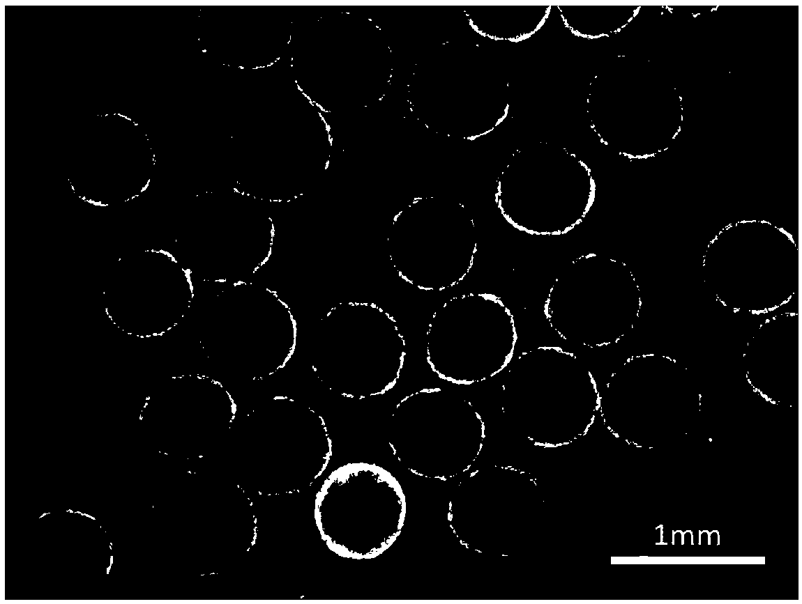
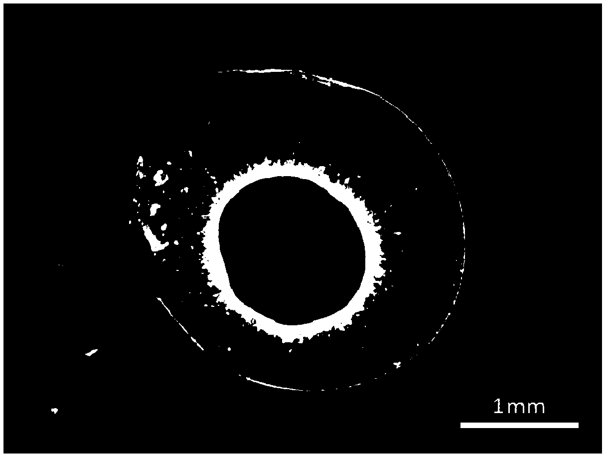
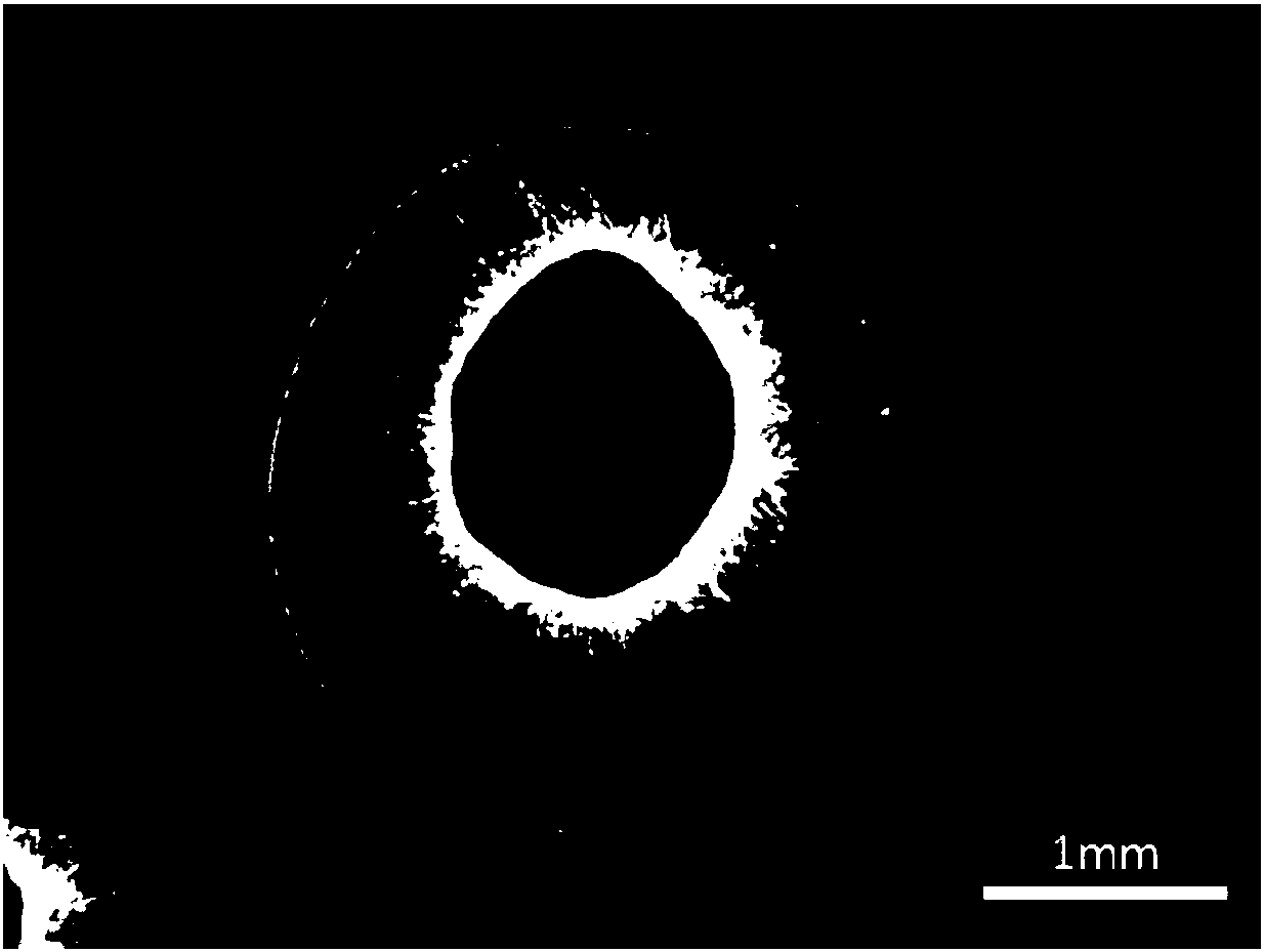
[0035] Step 1. After obtaining the neurosphere from the RONA method (see the article Cultured networks of excitatory projection neurons and inhibitory interneurons for studying human cortical neurotoxicity published by Xu JC and Fan J on Science Translational Medicine in April 2016), the second Days were digested with accutase into single cells. After counting, the same number of 5000 cells were inoculated into the wells of a 96-well cell culture plate with round bottom, and placed on a circular shaker in a carbon dioxide incubator at 37 degrees Celsius using medium A. Cultivate for 7 days, change the medium every 3 to 5 days; medium A is Neurobasal and B27 additives (without Vitamine A) containing final concentration of 2μM retinoic acid, 20ng / ml BDNF and GDNF, 0.2mM ascorbic acid, 10μM cAMP, Among them, the dosage ratio of ...


PUM

PropertyMeasurementUnit
diameteraaaaaaaaaa

Abstract

The invention provides a preparation method of a 3D (Three Dimensional) brain organ. The preparation method comprises the following steps: digesting neurospheres obtained by an RONA method into singlecells through accutase; after counting, inoculating a cell culture plate with the single cells and culturing in a culture medium A; after culturing to the 7th day, continually culturing in a culturemedium B; when culturing to the 25th to 35th day, covering the neurospheres with Matrigel; then continually culturing to the 55th to 65th day; after covering the neurospheres with the Matrigel for thesecond time, continually culturing. The invention further provides a culture medium for culturing the 3D brain organ. According to the preparation method provided by the invention, the highly-purified neurospheres obtained by the RONA method can be controlled and cultured by utilizing a relatively simple method to obtain the real 3D brain organ with uniform size and structure; the 3D brain organhas a six-layer cortical structure of a brain and each subtype of inhibitory medial nerve cells; the 3D brain organ is applicable to in-vitro disease researches, medicine screening and the like and has great industrialization significance.

Description

Technical field [0001] The invention belongs to the technical field of 3D brain organoids, and particularly relates to a method for preparing 3D brain organoids from human neurospheres. Background technique [0002] Human nerve cells or brain tissues are difficult to obtain, and they are almost impossible to culture in vitro, making many neurological disease research and drug development stagnant without a good human model. Human nerve cells derived from embryonic stem cells or induced pluripotent stem cells as models of neurological diseases have become an international innovation technology and hot spot in recent years. At the same time, because 3D culture can better simulate the brain tissue environment and cell communication, it has an important role in promoting the maturation and function of nerve cells, and the obtained 3D brain organoids have an irreplaceable role in the study of human brain development and related diseases. In vitro culture of various human 3D organs ha...

Claims


Application Information

Patent Timeline
Patent Type & Authority Applications(China)
IPC IPC(8): C12N5/079
CPCC12N5/0618C12N2500/38C12N2500/40C12N2501/13C12N2513/00C12N2533/54C12N2509/00C12N2523/00C12N2527/00
Inventor 范靖王安欣徐金翀邹潭
Owner ZHEJIANG HUODE BIOENG CO LTD
Why Eureka
Try Eureka